Sahar Hamed, Nashwa Barakat, Mohamed Sobh, Nahla Anber, Basma Hamed and Fatema Moustafa
Many apoptotic and fibrotic markers are known to be important in the development of renal fibrosis. In Unilateral Ureteral Obstruction (UUO) the Obstructed Kidney (OK) develops fibrosis, while the Contralateral (CL) does not. In this study we investigated the gene expression of different apoptotic and fibrotic molecules which may affect fibrosis developments due to UUO. UUO was prepared under isoflurane anesthesia, and the animals were sacrificed after 3, 7 and 14 days post UUO. UUO caused hydronephrosis, dilation of renal tubules, loss of parenchymal thickness, apoptosis and fibrosis. Damage was most severe in mice sacrificed after 14 days of UUO, while both 3 and 7 days groups showed considerably milder hydronephrosis, no tubular necrosis, and less tubular dilation. We detected increased levels of transforming growth factor β (TGF-β) and alpha-smooth muscle actin (α-SMA), Matrix metalloproteinase 2 (MMP2) and MMP9 (fibroblast activation marker) and TNF as apoptotic marker in the kidney tissue of UUO mice relative to the control UUO mice. This increase was confirmed by immunohistochemistry and gene expression as well. In conclusion, we found that the different pro-fibrotic molecules, α-SMA, TGF-β, MMP2, MMP9, Fibronectin and pro-apoptotic molecule, TNF expression were increased in UUO mouse kidney compared to contralateral or sham kidney. This increase was found to be time dependent.
Maruf-ul-Islam, Md Hasibul Hasan Joarder, Koushik Ahamed, Md Ben Yameen, Rokshana Sharmin, AHM Khurshid Alam and Ariful Islam
The aim of the study to investigate the effect of Aqueous Extract of B. ceiba Young Roots (AEBCYR) on the absorption of carbohydrates (mono-, di-, and poly-saccharide) from the intestine in healthy rats. Eighty healthy Long Evans rats were randomly assigned into eight groups (n=10 per group). The systemic absorption of carbohydrates was examined after loading a mixture of carbohydrates separately with AEBCYR in the ratio of 3:2 by using a glucometer. Furthermore, the sGPT and sGOT levels were determined by a semi bio-analyzer. Evaluations were based on a significance level of p<0.05. Aqueous extract of B. ceiba young roots (G-AEBCYR) solution inhibited glucose absorption up to 90 min and the glucose level was found to be 3.2 ± 0.22 mmol/L. Similarly, Suc- AEBCYR and Sta- AEBCYR solutions inhibited sucrose and starch absorption up to 120 and 180 min, and the reduction of glucose level was 39.22% and 56.37%, respectively, when compared with the standard CMC solution. On the other hand, the CMC mixed glucose (G-CMC), sucrose (Suc-CMC), and starch (Sta-CMC) solutions inhibited the maximum glucose absorption up to 60, 90, 120 min. Also, the AEBCYR reduced the hepatotoxicity by detecting the decreased levels of sGPT and sGOT compared to the CMC. Phytochemical analysis revealed the presence of fibers, alkaloid. Our results indicate that the AEBCYR delays carbohydrate absorption from the intestine of normal rats. It also showed a remarkable decrease of sGPT and sGOT levels, which indicates hepatoprotective activity.
Jun Ma, Chun-Xia Xiao, Kang-Jia Liu and Ren-Wang Jiang
Objective: To establish a method for quantitative determination of three quassinoids (bruceoside B, bruceoside A and brusatol) in Brucea javanica by HPLC.
Method: The determination was carried out on a Cosmosil (4.6×250 mm, 5 μm) C18 column eluted by a gradient elution program of H2O (A)-CH3OH (B) at a flow rate of 1.0 mL min-1, and the detection wavelength was 221 nm.
Result: The calibration curve of bruceoside B, bruceoside A and brusatol were linear in the range of 0.722-2.166, 2.074-6.222 and 0.503-1.509 μg, respectively with all the correlations of 0.9999. The average recovery of bruceoside B, bruceoside A and brusatol were 96.1%, 106.3% and 96.7%, with the RSD of 4.4%, 5.9% and 4.8% (n=5), respectively.
Conclusion: The results showed that the content of bruceoside B was in the range of 0.05%~0.12%, bruceoside A was in the range of 0.19%~0.38% and brusatol was in the range of 0.07%~0.18%, indicating that the contents of bruceoside B, bruceoside A and brusatol varied among different sources. Furthermore, the fingerprinting chromatograms of eight samples of Brucea javanica were established and the characteristic peaks were identified by mass spectrometry. The method was accurate, simple and convenient, which could be used for the quality control of Brucea javanica.
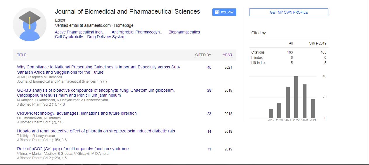
Abdullahi Umar Ibrahim, Mehmet ÖZSÖZ, Zubaida Saeed, Galaya Tirah and Ojo Gideon
CRISPR technology is the most widely used genetic editing tool to edit, modify, delete, add and replace DNA sequences in both prokaryote and eukaryote, the normal CRISPR pathway cut gene at a specific point by recognizing a Protospacer Adjacent Motif (PAM) and induce double strand break, the cell can repair this destruction through Non- Homologous Enjoining. Scientist takes advantage of this break and inserts a homologous strand with specific changes through Homologous Directed Repair (HDR). CRISPR Interference (CRISPRi) follow the same technique but use a dead, mutated or inactive form Cas9 protein to render gene inactive and thereby block gene expression. In this study we compare both the similarities between CRISPR-Cas9 system and CRISPR Interference and further shown the differences of off-target in both systems.
Recruitment of Microcirculatory-Mitochondrial (RMM) reduces Microcirculatory-Mitochondrial Distress Syndrome (MMDs), and Syndrome of Multi-Organ Dysfunction (MODs), by accelerated speed of delivery and return of blood flow which directly leads to a decrease in tissue hypoxia marker pCO2 (AV gap) and respectively with ↓ many other Endogenous Toxic Substances (ETS).
In cases of pulmonary damage with ↑ pCO2 & ↓ Oxygenation Index PaO2/FiO2 ↓ 300 the development of Acute Respiratory Distress Syndrome (ARDs), MMDs are also aggravated at ↑ with pCO2 AV gap. RMM also needs additional support of Multiple Organ Therapies-Multi-Organ Supportive Therapy (MOST), Alveolar Recruitment, Extracorporeal Life Support Organization (ELSO), Modeling of the Index of Extravascular Lung Fluid, EVLWI, Th4- Th5 Thoracic Epidural Block, Active detoxification methods.
The absence of decreasing of the pCO2 tissue hypoxia marker at the pCO2 AV gap ↓ 5.0 mmHg, after RMM proves the mitochondrial eu-energetic metabolic remodeling with the elimination of the hypo(an)ergic mitochondria performed by lysosomal clearance (mitophagy) makes the predominance eu-ergic mitochondria with the normalization of mitochondrial Ca++-uniporter-channel and mitochondrial permeability pore transition which productively inactivate the toxic forms of oxygen and nitrogen.

 Spanish
  Chinese
  Russian
  German
  French
  Japanese
  Portuguese
  Hindi
Hindi 