Tolga Suvar*, Jill Mhyre and Nadir Sharawi
Purpose: The purpose of this case report is to demonstrate the management of a parturient with a life-threatening pulmonary embolism in the antenatal and perinatal phase of her care. In addition to this, it is important to highlight cardiopulmonary resuscitation which was titrated by life radiological imaging during rescue catheter embolectomy. By observing these images, the medical team appreciated the progressive hypokinesis of the myocardium and thus anticipating prompt and high-quality chest compressions.
Clinical Features: A 24-year-old woman at 37 weeks and 4 days gestation who was transferred from an outside hospital on a heparin infusion to our University Medical Center for peripartum care and management of a suspected pulmonary embolism (PE). On admission she was extremely tachycardic and short of breath. She underwent an emergent cesarean delivery at the mother’s request for category 3 non-reassuring fetal heart tones. After induction of general anesthesia, she became profoundly hypotensive despite ongoing fluid resuscitation and vasopressor support. A transesophageal echo (TEE) revealed a dilated right ventricle with severe right heart strain. Inhaled nitric oxide and a milrinone infusion were started due to cardiogenic shock. The decision was made to transfer her to the interventional radiology (IR) suite for an emergent pulmonary angiography and a rescue catheter embolectomy. While performing the embolectomy in the interventional radiology suite, the patient suffered three episodes of cardiac arrest. During continuous x-ray screening we were able to appreciate real-time images of the heart during the full cardiac cycle, the right ventricle appeared visibly dilated and hypokinetic. Contractility continued to progressively get worse up until the point of cardiac arrest after each event. We were able to guide further therapeutic decisions (ongoing fluid therapy, vasopressor, and inotropic support) based on the real-time fluoroscopy images that were visualized. Continuous images during this period of hemodynamic instability were captured along with the images of the thrombectomy catheter removing large amounts of embolic thrombus from both pulmonary arteries.
Conclusion: Patients who are undergoing interventional radiological procedures are often too sick to undergo major surgery and in this scenario the utility of live x-ray was paramount to the quality of chest compressions and thus survival of this patient 1.
HTML PDFShare this article
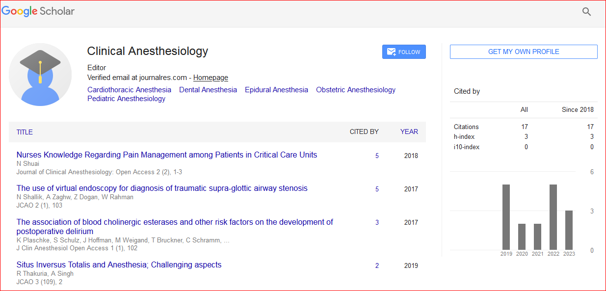
Journal of Clinical Anesthesiology: Open Access received 31 citations as per Google Scholar report