Case Report - (2020) Volume 4, Issue 1
Received: 16-Apr-2020
Published:
30-Apr-2020
Citation: Tolga Suvar , Jill Mhyre and Nadir Sharawi. "Life
Threatening Pulmonary Embolism During Pregnancy Requiring Emergency
Embolectomy ". JCAO, an open access journal 4 (2020): 113.
Copyright: © 2020 Suvar T, et al. which permits unrestricted use, distribution and reproduction in any medium, provided the original author and source are credited.
Purpose: The purpose of this case report is to demonstrate the management of a parturient with a life-threatening pulmonary embolism in the antenatal and perinatal phase of her care. In addition to this, it is important to highlight cardiopulmonary resuscitation which was titrated by life radiological imaging during rescue catheter embolectomy. By observing these images, the medical team appreciated the progressive hypokinesis of the myocardium and thus anticipating prompt and high-quality chest compressions.
Clinical Features: A 24-year-old woman at 37 weeks and 4 days gestation who was transferred from an outside hospital on a heparin infusion to our University Medical Center for peripartum care and management of a suspected pulmonary embolism (PE). On admission she was extremely tachycardic and short of breath. She underwent an emergent cesarean delivery at the mother’s request for category 3 non-reassuring fetal heart tones. After induction of general anesthesia, she became profoundly hypotensive despite ongoing fluid resuscitation and vasopressor support. A transesophageal echo (TEE) revealed a dilated right ventricle with severe right heart strain. Inhaled nitric oxide and a milrinone infusion were started due to cardiogenic shock. The decision was made to transfer her to the interventional radiology (IR) suite for an emergent pulmonary angiography and a rescue catheter embolectomy. While performing the embolectomy in the interventional radiology suite, the patient suffered three episodes of cardiac arrest. During continuous x-ray screening we were able to appreciate real-time images of the heart during the full cardiac cycle, the right ventricle appeared visibly dilated and hypokinetic. Contractility continued to progressively get worse up until the point of cardiac arrest after each event. We were able to guide further therapeutic decisions (ongoing fluid therapy, vasopressor, and inotropic support) based on the real-time fluoroscopy images that were visualized. Continuous images during this period of hemodynamic instability were captured along with the images of the thrombectomy catheter removing large amounts of embolic thrombus from both pulmonary arteries.
Conclusion: Patients who are undergoing interventional radiological procedures are often too sick to undergo major surgery and in this scenario the utility of live x-ray was paramount to the quality of chest compressions and thus survival of this patient 1.
On admission, our patient was extremely tachycardic (HR 150 bpm) and short of breath (RR 35/min) (Figure 1). The fetal tracing demonstrated category 3 non-reassuring fetal heart tones which did not resolve despite intrauterine resuscitation [1]. After a concise review of the treatment options among the multidisciplinary team, we recommended to the patient that she continue anticoagulation therapy with a heparin infusion, despite the very real risk that her fetus would not survive. Due to her underlying critical illness general anesthesia and cesarean delivery were considered not to be in the best interests of the mother. After careful consideration of the options the patient desired emergent cesarean delivery to minimize the risk of fetal demise with the knowledge that she may die in the process either from her underlying medical condition or from postpartum hemorrhage secondary to anticoagulation.
After induction of general anesthesia, she became profoundly hypotensive despite ongoing fluid resuscitation [2]. A norepinephrine infusion was commenced and a transesophageal echo (TEE) revealed a dilated right ventricle with severe right heart strain [3]. Inhaled nitric oxide and a milrinone infusion were started due to cardiogenic shock. A live male infant was delivered with APGAR scores of seven and eight at one and five minutes respectively. She remained hemodynamically unstable and her circulation required further support with an infusion of norepinephrine and vasopressin. Her condition continued to deteriorate despite maximal medical therapy [4].

Figure 1. EKG tracing displays sinus tachycardia with S1Q3T3 pattern
The decision was made to transfer her to the interventional radiology (IR) suite for an emergent pulmonary angiography and a rescue catheter embolectomy (Figures 2,3). During the procedure she suffered three episodes of pulseless electrical activity (PEA) cardiac arrest.
Cardiopulmonary resuscitation (CPR) was successful after every event of cardiac arrest [5,6].

Figure 2. CTA of chest displaying bilateral blood clots in pulmonary arteries

Figure 3. Interventional radiology snapshot of filling defects from the contrast filled pulmonary vasculature
Due to the small confines of the IR suite, the location of the anesthetic machine, and the “C-arm” along with the requirement to try and maintain a sterile surgical field, access to the patient was remarkably difficult. Invasive cardiac output monitoring or TEE was not practical in this environment [7,8].
This case is unique in that it became apparent during continuous x-ray screening, that we were able to appreciate real-time images of the heart during the full cardiac cycle. The right ventricle appeared visibly dilated and hypokinetic. Contractility continued to progressively get worse up until the point of cardiac arrest after each event. We realized that we were able to guide further therapeutic decisions (ongoing fluid therapy, vasopressor, and inotropic support) based on the live images that we visualized. Continuous images during this period of hemodynamic instability were captured along with the images of the thrombectomy catheter removing large amounts of embolic thrombus from both pulmonary arteries [9].
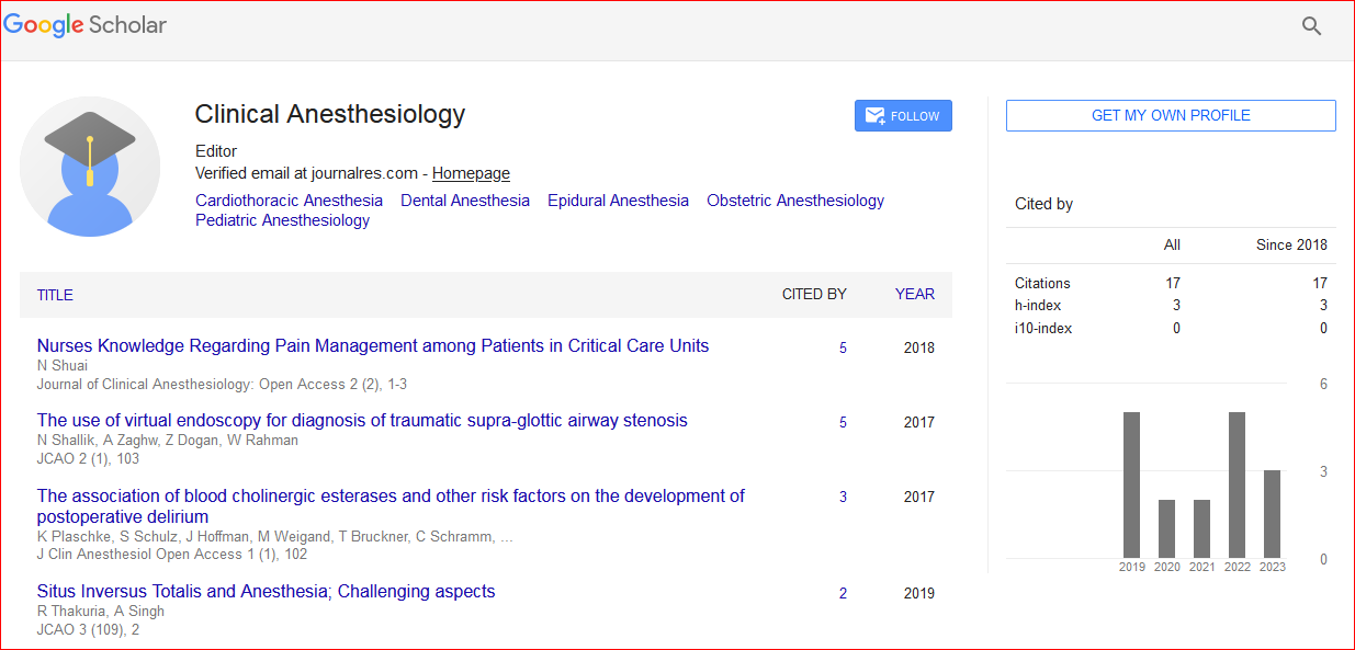
Cumulative CPR time was estimated to be approximately twenty-five minutes. The procedure was successful and she continued to improve in the intensive care unit. She was discharged from hospital on a postoperative day five with no signs of gross neurological deficit. At a five-month follow-up, thrombophilia screen was negative and the patient is back in full-time employment as a critical care nurse.
Journal of Clinical Anesthesiology: Open Access received 31 citations as per Google Scholar report