Amer A Taqa
The point of this investigation was to decide the compound attribute of outstanding dentine after caries evacuation with either test chemo mechanical caries expulsion specialist (ECMCRA) or Cariso® Twenty extricated human third molars were installed independently in moderate restoring epoxy pitch; the teeth were decoronated and ground level the occlusal surfaces. A 280 ± 20 µm thick layer of incompletely demineralized dentine was made on the occlusal dentine surface by pH cycling. From that point forward, the teeth conveyed to two gatherings and separated longitudinally. The principal half caries was unearthed, and a flimsy layer of dentine was tenderly scratched off with careful surgical tool sharp edge and exposed to FTIR investigation which is considered as a leftover dentine with no CMCR specialists. The subsequent parts were gotten after carious tissues evacuation with either ECMCRA or Cariso (MediTeam Dental. Sweden) individually as per the makes directions and this is considered as a leftover dentine after caries evacuation with specialists. The FTIR spectra of each example were acquired by ALPHA FTIR spectrometers (Bruker, Germany) with 4.0 cm goal, with the scope of 500-4000 cm. To assess the trustworthiness of the collagen triple helix, top absorbance proportions of 1235 cm/1454 cm were thought of.
No vanishing or move of groups was apparent about the mineral and natural substance of residual dentine. ECMCRA did not advance collagen denaturation.
The synthetic investigations in the current examination for the leftover dentine after caries expulsion with either ECMCRA or Cariso infer that excess dentine after evacuation with strategies for CMCR are irrelevantly contrast from control dentine. It is notable that caries attack prompts the separation of dentin into zones with modified synthesis, collagen uprightness and mineral personality. Notwithstanding, comprehension of these progressions from the principal viewpoint of sub-atomic structure has been missing up until now. Considering this, the current work means to use Fourier change infrared spectroscopy (FTIR) to straightforwardly separate atomic data with respect to collagen's and hydroxyapatite's primary changes as dentin advances from the straightforward zone (TZ) into the ordinary zone (NZ). Unembedded ultrathin dentin films were segmented from carious teeth, and a FTIR imaging framework was utilized to get spatially settled FTIR spectra.
As indicated by the mineral-to-network proportion picture created from enormous territory low-unearthly goal filter, the TZ, the NZ and the middle of the road sub transparent zone (STZ) were distinguished. High-ghastly goal spectra were taken from each zone and in this manner analysed concerning mineral substance, carbonate circulation, collagen denaturation and carbonate replacement designs. The honesty of collagen's triple helical structure was additionally assessed dependent on spectra gathered from demineralized dentin movies of chose teeth. The outcomes uphold the contention that STZ is the genuine sclerotic layer, and they prove the set-up information that collagen in TZ is not really changed and subsequently should be saved for reparative purposes. Also, the nearby likeness between the STZ and the NZ regarding carbonate content, and that between the STZ and the TZ as far as being A-type carbonate-rich, propose that the mineral that at first blocks dentin tubules is hydroxyapatite recently produced from odontoblastic exercises, which is then changed into whitlockite in the demineralization/remineralization measure as caries advances. Human molars with occlusal carious sores were gathered from the Oral Surgery Clinic at the University of Missouri-Kansas City (UMKC) School of Dentistry in the wake of acquiring the patients' educated assent under a convention endorsed by the UMKC grown-up wellbeing sciences institutional audit board.
Removed teeth were put away at 4°C in 0.96% (w/v) phosphate-supported saline containing 0.002% sodium azide prior to being part fifty-fifty in the occlusal-apical course through the injuries utilizing a moderate speed water-cooled jewel saw (Buehler, Lake Bluff, Ill., USA). A sum of 7 teeth were chosen (assigned T1–T7), all of which had half or a greater amount of the cross-sectional territory being evidently typical dentin under a light magnifying lens and were in this way exposed to additional preparing.
During the microtubing cycle, the tooth tests and the blade edge were kept soggy with refined water, and the segmented dentin films were permitted to slide onto the blade and later gathered on 13-mm-width, 1-mm-thick barium fluoride (BaF2) plates with a fine paintbrush. For every tooth, its relating unpermineralized dentin films were investigated under a light magnifying instrument, from which one of the most un-broke was picked to speak to the tooth and continue with FTIR imaging.
PDFShare this article
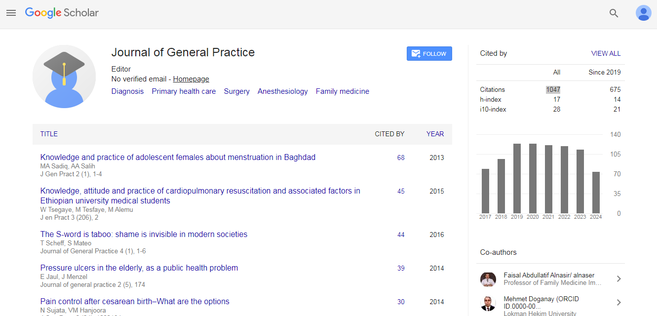
Journal of General Practice received 1047 citations as per Google Scholar report