Research Article
Pages: 1 - 4Ramesh Ramji, Ang Chee Xiang, Ng Jia Ying, Lim Chwee Teck and Chen Chia Hung
One of the essential issues in cell biology is to analyze intracellular proteins that play an important role in signal transduction and processes. Standard assays for intracellular proteins are based on ensemble measurements which rule out the differences of intracellular protein expression in individual cells. Especially in cancer biology, only a few tumour cells turn into circulating tumour cells causing metastasis. In such cases, understanding the heterogeneity in cellular physiology would be necessary. To screen for intracellular protein expression among individual cells effectively, a reliable method to lyse individual cells in a high throughput manner is essential. Here, we demonstrate an integrated microfluidic platform combining a cell focusing channel, reagent injection inlet and a droplet generator for high throughput single cell lysis. Individual cells are encapsulated into the droplets with chemical reagents which result in disrupting the cell membrane integrity, causing cell lysis. High throughput mammalian cell lysis was performed thus facilitating intracellular protein analysis on a micro device
Research Article
Pages: 1 - 5Aminata P. Kilungo, Njeri Carlton-Carew and Linda S. Powers
With the rapid increase in global population, geographically changing drought conditions and the ensuing potential water shortage, water quality has become a major concern. In some extreme cases, such as Arizona, the population may have to switch and use recycled toilet water for potable use in the near future. However, our current monitoring methods for drinking water do not provide fast and reliable results to deal with these challenges. By using intrinsic fluorescence, microbial contamination in water can be monitored in real-time, continuously, without sample collection or contact and at very low concentration. The detection limit of the instrument designed specifically for this purpose and reported here is ~50 bacterial cells/L. By monitoring the fluorescence of cellular components of microorganisms, their concentrations and metabolic states (live, dead, spores) can be determined. These fluorophores include reduced pyridine nucleotides (RPNs), flavins, and cytochromes to distinguish live cells; cytochromes for dead cells; and calcium dipicolinic acid (DPA) for spores. By using this method, a wide range of microorganisms such as bacteria, protozoa, amoebae, fungi and other microorganisms can be detected
Research Article
Pages: 1 - 4Waqas Saleem and Patricia A Broderick
Here, we present results from two independent studies carried out using Neuromolecular Imaging (NMI) with miniature BRODERICK PROBE ® biosensors. In the first study, we imaged neurotransmitters and neurochemicals in human epilepsy patients intraoperatively during early and late neurodegeneration. In the second study, we imaged neurotransmitters and neurochemicals in an experimental murine model using animals with and without neurodegeneration caused by Parkinson’s disease (PD). We compared our results derived from animals with lesioned group (PD) with non-lesioned group (non-PD), using the same in vivo NMI paradigm. NMI biotechnology enabled neurotransmitters, neuropeptides and neurochemical imaging of dopamine (DA), serotonin (5-HT), homovanillic acid (HVA), L-tryptophan (L-TP), dynorphin A (DYN A) and somatostatin (SRIF). Each neurotransmitter and neurochemical was imaged at its respective signature i.e., its electroactive oxidation/half-wave potential. Results showed neuropeptide signatures of DYN A and SRIF as common biomarker molecules following late neurodegeneration in epilepsy patients and in PD animal models. Placing these two studies together allowed us to us to provide a new hypothesis about a possible biomarker link between the two neurodegenerative diseases, epilepsy and PD. Interestingly, this biomarker link, to our knowledge has not been observed previously. These findings will provide new strategies for better diagnoses, detection of and protection against epilepsy and Parkinson’s disease
Research Article
Pages: 1 - 7Ugur Korcan Demirok, Aman Verma, and Jeffrey T La Belle
With an increasing global population, rising healthcare costs, and greater demand on hospitals and clinicians, a growing need for low cost, rapid, Point of Care Technologies (POCT) exists. The overall goal is to detect or monitor a disease in order to give patients and clinicians fast, accurate, and all-encompassing information regarding the state of the disease. A challenge for many currentpoint-of-care technologies is the difficulty of monitoring several biomarkers simultaneously without the complexity of multiple sensors, labels, or spatially separated transducers. We have previously shown that convoluted signals obtained from protein biomarkers monitored by electrochemical impedance spectroscopy can be “tuned” away from one another by conjugation with gold nanoparticles to allow for the potential simultaneous detection of multiple biomarkers. This method of detection yields a sensitive and specific means of biomarker quantification in human media including tears or blood. In this work, we detail the development of a mathematical model that explores the roles of various factors, such as nanoparticle size and the nature of materials, so that a design space could be created for tuningotherwise convoluted biomarkers. Furthermore, we present assessments as to the validity of this model with preliminary bench-top experiments by taking advantage of gold nanoparticle-antibody conjugates of varying sizes. Gold nanoparticle size changes of 5, 10, and 20 nm demonstrated a 10.0, 4.8, and 1.0 Hz shift in frequency, respectively. Future work includes exploration of different sensor configurations, continuous monitoring, and the prospect for implantable sensors were also discussed as potential future avenues.
Research Article
Pages: 1 - 7Vinay Bhardwaj, Supriya Srinivasan and Anthony J McGoron
Among several physical, chemical and immunoassay-based methods for the detection of biomolecules, the Enzyme-Linked Immuno-Sorbent Assay (ELISA) is the standard technique that is routinely used for quantification of known proteins. However, it is a label-based, end-point sensor technique that is time-consuming, labor-intensive and fairly costly. This sandwich assay typically involves a series of peptide binding and washing steps. Here, we report a Surface-Enhanced Raman Spectroscopy (SERS) immuno-nanosensor technique that allows rapid and label-free extracellular detection of proteins compared to ELISA, and can potentially be used for intracellular detection. Our study shows that the silver nanoparticles (AgNPs) based SERS sensor can detect the stress-proteins, HSP70 and RAD54 expressed by yeast in response to environmental-toxins, in a dose dependent manner. As compared to the multi-step sandwich ELISA technique, the detection of stress-proteins using the SERS sensor is a simple two-step process. The simplicity of the SERS nanosensor design allowed the rapid detection of proteins within two hours in a fairly cost-effective and user-friendly approach. The SERS sensor we reported has an edge over ELISA as it directly quantifies the proteins without using any label (label-free) and also gives qualitative information about the antigen-antibody interaction. The SERS sensor showed good correlation and comparable sensitivity with ELISA. However, SERS was found to be less reproducible. Compared to previous reports on SERS-based protein detection techniques, our colloidal SERS sensor is easy to fabricate, offers improved biocompatibility, and allows rapid detection of the proteins in a cellular environment at picogram-levels. As a result, the SERS sensor demonstrates great potential for biomedical and environmental sensor technology (BEST) allowing label-free, rapid and sensitive detection, and could possibly replace ELISA.
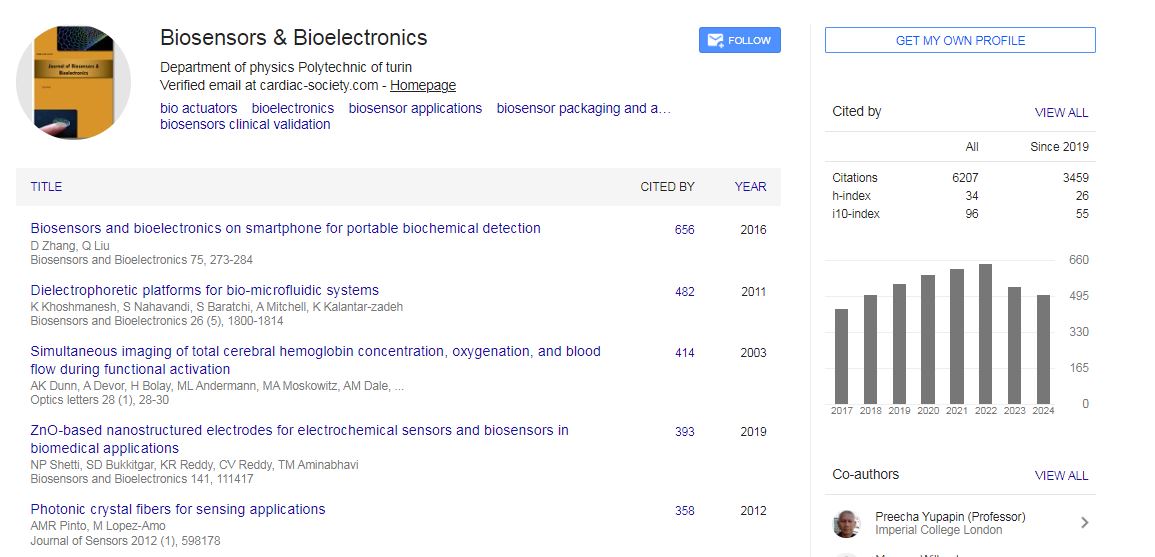
Biosensors & Bioelectronics received 6207 citations as per Google Scholar report
 Spanish
  Chinese
  Russian
  German
  French
  Japanese
  Portuguese
  Hindi
Hindi 