Editorial - (2022) Volume 10, Issue 2
Received: 01-Mar-2022, Manuscript No. Jcmg-22-68498;
Editor assigned: 03-Mar-2022, Pre QC No. P-68498;
Reviewed: 07-Mar-2022, QC No. Q-68498;
Revised: 12-Mar-2022, Manuscript No. R-68498;
Published:
18-Mar-2022
, DOI: 10.37421/2472-128X.2022.10.201
Citation: Nath, Sanjay. “Radiogenomics for Cancer.” J Clin Med Genomics 10 (2022): 201.
Copyright: © 2022 Nath S. This is an open-access article distributed under the terms of the Creative Commons Attribution License, which permits unrestricted use, distribution, and reproduction in any medium, provided the original author and source are credited.
Radiogenomics, a mix of "Radiomics" and "Genomics," utilizing Artificial Intelligence has as of late arisen as the best in class science in accuracy medication, particularly in oncology care. Radiogenomics organizations huge scope quantifiable information removed from radiological clinical pictures wrapped with customized genomic aggregates. It manufactures an expectation model through different AI strategies to define the gamble of patients, screen remedial methodologies, and evaluate clinical results. It has as of late shown enormous accomplishments in anticipation, therapy arranging, endurance expectation, heterogeneity examination, reoccurrence, and movement free endurance for human disease study. Despite the fact that AI has shown huge execution in oncology care in different clinical viewpoints, it has a few difficulties and limits. The proposed audit furnishes an outline of radiogenomics with the perspectives on the job of AI as far as its commitments for computational as well as oncological viewpoints and offers accomplishments and amazing open doors in the period of accuracy medication.
Malignant growth is a subsequent driving reason for death around the world, just after cardiovascular illnesses, representing almost 10 million passings in 2020. According to world wellbeing association (WHO) insights, the normal sorts of malignant growths that individuals experience more are those of the bosom, lungs, colorectal, prostate, skin, cerebrum, and stomach. The disease trouble keeps on developing universally, applying enormous physical, close to home, and monetary stress on people, families, networks, and wellbeing frameworks. Nations with unremarkable and chronic weakness framework don't have the admittance to convenient, quality conclusion and therapy for countless patients.
In the period of accuracy medication, atomic portrayal of malignant growth utilizing genomic innovation is fundamental. Over the most recent couple of years, critical advancement has been seen in atomic portrayal. In any case, because of the specialized intricacy, cost, and completion time, an immense scope genome-based portrayal of malignant growth isn't yet regularly adjusted for a wide range of diseases. In existing clinical practices, because of the heterogeneous way of behaving of malignant growth, sub-atomic profiling is much of the time restricted, and heterogeneity of the growth is over and over missed when a piece of the disease is inspected . All through the treatment, assurance of atomic targets requires ex vivo postoperative investigation of the resected cancer or biopsy test [1,2]. This has limited the appraisal of growths' spatial and fleeting heterogeneity and is beyond the realm of possibilities to expect to decide the sub-atomic change of disease constantly. Furthermore, on account of the strong kind of cancers, the practical, anatomic, and physiological properties of the entire growth may not be completely reflected in the histopathological tests. Specialists and researchers overall have seen the significant occupation of clinical imaging in clinical therapy navigation and in dissecting diseases. Prior, its principal work was limited to anticipation and arranging . In any case, as of late, imaging-got markers acquired from clinical pictures have altogether been examined to convey knowledge into disease painlessly. In particular, imaging portrays the peritumoral locales, as these districts are not consistently obtrusively taken out for the sub-atomic portrayal of disease [3-5].
None.
Google Scholar, Indexed at, Crossref
Google Scholar, Indexed at, Crossref
Google Scholar, Indexed at, Crossref
Google Scholar, Indexed At, Crossref
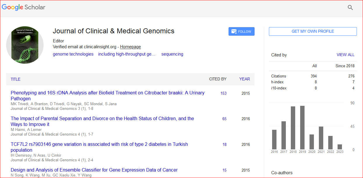
Journal of Clinical & Medical Genomics received 391 citations as per Google Scholar report