Perspective - (2022) Volume 6, Issue 1
Received: 03-Jan-2022, Manuscript No. hps-22-64474;
Editor assigned: 04-Jan-2022, Pre QC No. P-64474;
Reviewed: 13-Jan-2022, QC No. Q-64474;
Revised: 18-Jan-2022, Manuscript No. R-64474;
Published:
25-Jan-2022
, DOI: 10.37421/2573-4563.2022.6.180
Citation: Danielle, Flavia. “Pancreatic Disease Diagnosing in Patients.” Hepatol Pancreat Sci 6 (2022): 180.
Copyright: © 2022 Danielle F. This is an open-access article distributed under the terms of the Creative Commons Attribution License, which permits unrestricted use, distribution, and reproduction in any medium, provided the original author and source are credited.
Acute and chronic inflammatory disorders, as well as neoplastic malignancies, affect the pancreas. Because biochemical and imaging signals may be unspecific and only appear at an advanced stage of the disease, it is difficult to diagnose these patients early. Pancreatic imaging advancements are critical for early diagnosis of pancreatic disorders. Over the last decade, ultrasonography has seen significant technological advancements as a diagnostic tool. It is still the primary method for first-line imaging, and it is increasingly being used to enhance decision-making by clarifying data from other imaging modalities [1].
Chronic Pancreatitis (CP) causes irreversible pancreatic parenchymal and ductal alterations. Magnetic resonance imaging (MRI) may be able to offer an early diagnosis of CP, allowing people to be treated or given treatment alternatives that will help them avoid progression. The presence of clinical symptoms, pancreatic exocrine function testing (the "gold" standard), and imaging are commonly used to diagnose early CP. MRI can be used to diagnose CP by examining both parenchymal and ductal alterations, which is especially useful in individuals with advanced CP. The most common way to confirm the diagnosis is to check serum amylase and lipase levels. Although hyperamylasemia is prevalent in the majority of patients with acute pancreatitis, it can also be associated with other non-pancreatic acute abdominal disorders [2].
CT scanning confirms clinical and laboratory results with precision and gives excellent anatomic and morphologic representation of the pancreas and peripancreatic tissue. In the diagnostic workup of abdominal illnesses, transabdominal ultrasonography is still the most commonly utilised imaging modality. Ultrasonography is non-invasive, widely available, inexpensive, safe, and simple to use on a daily basis if necessary. Transabdominal ultrasonography, as a "real-time" imaging method, provides a comprehensive overview before localising the "area of interest" to undertake detailed examination and eventually pinpoint the origin of the condition. Acute pancreatitis is diagnosed by acute onset stomach pain and tenderness, primarily in the upper abdomen, increased pancreatic enzymes in the blood and/or urine, and pancreatitis features seen by diagnostic imaging such as ultrasonography (US) and CT [3-5].
Other stomach problems should be ruled out. After an acute pancreatitis diagnosis has been made, the aetiology should be determined in order to determine treatment options for acute pancreatitis or to prevent recurrence. Major arterial bleeding is a rare but significant pancreatitis consequence with a high morbidity and fatality rate. The gold standard for detecting a visceral artery pseudoaneurysm or the site of current bleeding in patients with pancreatitis has long been digital subtraction angiography (DSA). In comparison to DSA, multisection CT angiography is a minimally invasive technique that can generate high-resolution and high-contrast pictures of the artery lumen and wall with a substantially lower risk of complication and morbidity. Endoscopic stenting is the standard first-line treatment for individuals with locally progressed or metastatic illness who are jaundiced. Gastric outlet blockage is a common complication of pancreatic cancer, which affects 10–20 percent of people. Surgical bypass or endoscopic SEMS implantation can be used to provide relief. Patients with severe intractable abdomen or back pain may require increased analgesics, as described by the WHO analgesic ladder, as well as access to palliative care.
Indexed at, Google Scholar, Crossref
Indexed at, Google Scholar, Crossref
Indexed at, Google Scholar, Crossref
Indexed at, Google Scholar, Crossref
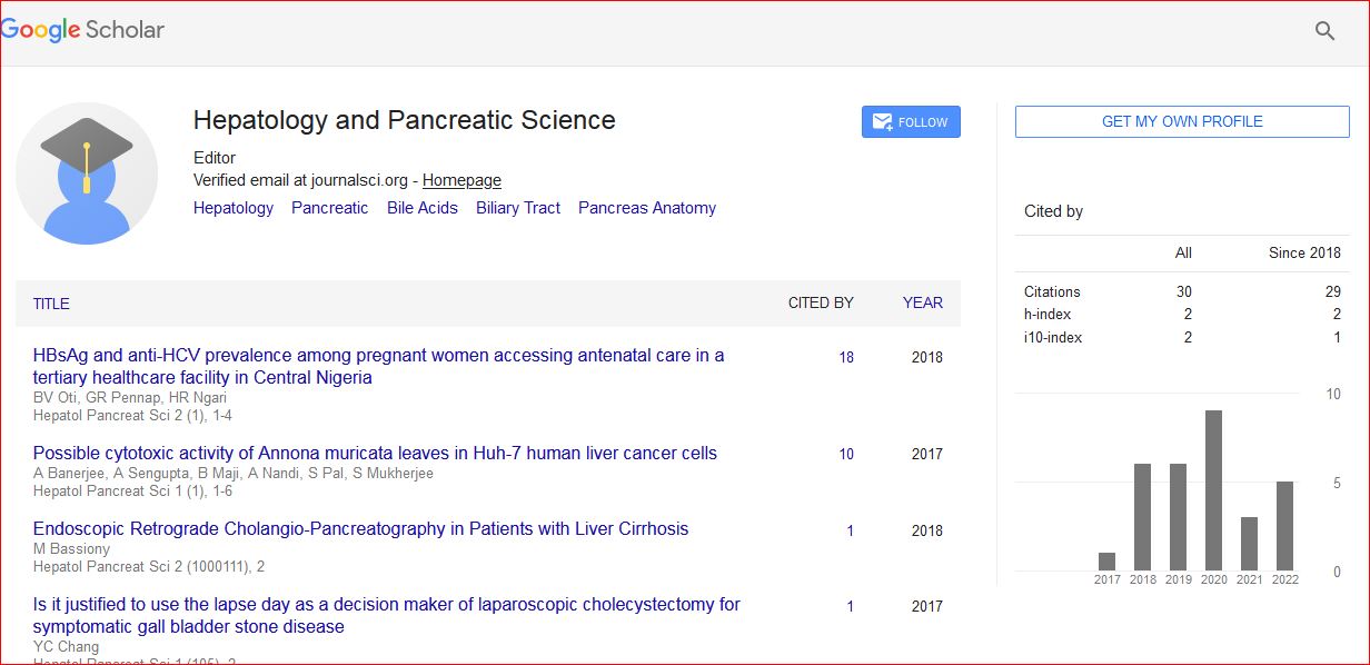
Hepatology and Pancreatic Science received 34 citations as per Google Scholar report