Brief Report - (2025) Volume 14, Issue 6
Received: 31-Oct-2025, Manuscript No. jmmd-26-184708;
Editor assigned: 03-Nov-2025, Pre QC No. P-184708;
Reviewed: 17-Nov-2025, QC No. Q-184708;
Revised: 21-Nov-2025, Manuscript No. R-184708;
Published:
28-Nov-2025
, DOI: 10.37421/2161-0703.2025.14.552
Citation: Popescu, Victor. ”Optimizing Malaria Diagnosis: Integrating Microscopy and RDTs.” J Med Microb Diagn 14 (2025):552.
Copyright: © 2025 Popescu V. This is an open-access article distributed under the terms of the Creative Commons Attribution License, which permits unrestricted use, distribution and reproduction in any medium, provided the original author and source are credited.
Malaria diagnosis is a critical component of effective control and elimination strategies, with microscopy historically serving as the gold standard due to its ability to identify parasite species and assess parasite density, crucial for clinical management [1].
However, the effectiveness of microscopy is often hampered by the need for trained personnel and quality-controlled equipment, limiting its utility in resource-constrained settings [1].
In parallel, rapid diagnostic tests (RDTs) have emerged as a faster, more accessible alternative, capable of detecting Plasmodium falciparum antigens with high sensitivity and specificity, making them invaluable for prompt diagnosis and treatment initiation, particularly in remote areas [1].
Despite their advantages, RDTs can suffer from false positives and negatives due to various factors, and they often cannot distinguish between viable and non-viable parasites or determine parasite density [1].
The choice between microscopy and RDTs therefore depends on local context, resource availability, and specific diagnostic needs, with a growing trend towards integrating both methods to optimize malaria case management [1].
The advent of malaria RDTs has revolutionized field diagnostics, offering unprecedented speed and ease of use compared to traditional microscopy, particularly beneficial in remote and resource-limited settings where trained microscopists and reliable equipment are scarce [2].
Their ability to provide results within minutes facilitates immediate treatment decisions, thereby contributing to reduced morbidity and mortality [2].
However, challenges remain, including the detection of low-density parasitemia and differentiation of Plasmodium species, especially non-falciparum malaria, necessitating careful quality assurance and training protocols to ensure accurate diagnosis and effective malaria control strategies [2].
Microscopy, while labor-intensive, offers a qualitative and quantitative assessment of malaria parasites in blood smears, allowing for species identification and parasite density estimation, vital for guiding treatment decisions, particularly for severe malaria cases [3].
Challenges with microscopy include the requirement for skilled personnel, strict quality control measures, and susceptibility to human error [3].
In contrast, malaria RDTs provide a rapid, antigen-based detection of malaria parasites, simplifying diagnosis in resource-limited settings, though their performance with other Plasmodium species can be variable and they generally do not provide parasite density information [3].
The integration of both methods, leveraging the strengths of each, is increasingly recognized as the optimal approach for comprehensive malaria diagnosis and surveillance [3].
The diagnostic landscape for malaria has evolved significantly, with both microscopy and rapid diagnostic tests (RDTs) playing critical roles [4].
Microscopy, a traditional method, offers detailed parasite information, including species identification and density, invaluable for assessing disease severity and treatment response, but demands skilled microscopists and robust quality assurance systems, limiting its application in remote areas [4].
RDTs have emerged as a vital tool for decentralized malaria diagnosis, providing quick results that enable prompt treatment and are highly sensitive for Plasmodium falciparum but may have limitations with low parasitemia or other Plasmodium species [4].
Understanding the strengths and weaknesses of each diagnostic modality is key to optimizing malaria control programs, with combined approaches often yielding the best outcomes [4].
The choice between malaria microscopy and rapid diagnostic tests (RDTs) is often dictated by local infrastructure, resource availability, and diagnostic needs [5].
Microscopy provides a comprehensive analysis, including parasite species and density, crucial for clinical decision-making, particularly for severe malaria, with its main limitations being the requirement for trained personnel and consistent quality control [5].
RDTs, on the other hand, offer speed and accessibility, making them ideal for primary healthcare settings and mass screening campaigns, generally reliable for detecting Plasmodium falciparum but may struggle with differentiating species or quantifying parasite burden [5].
Evaluating the performance of both in diverse epidemiological settings is essential to refine diagnostic algorithms and improve malaria control efforts [5].
Malaria diagnosis hinges on two primary methods: microscopy and rapid diagnostic tests (RDTs) [6].
Microscopy excels at identifying parasite species, quantifying parasitemia, and assessing morphological characteristics, providing a detailed diagnostic picture indispensable for research and managing complex cases, though its practical implementation is hindered by the need for skilled microscopists and consistent laboratory conditions [6].
RDTs offer a rapid, user-friendly alternative, especially in point-of-care settings, facilitating immediate treatment initiation, yet their performance can vary for other species and in cases of low parasite load, and their inability to determine parasite density limits their utility in severe malaria assessment [6].
Consequently, a balanced approach, often integrating both, is crucial for comprehensive malaria diagnosis [6].
The efficacy of malaria control programs is intrinsically linked to accurate and timely diagnosis [7].
Microscopy, the historical gold standard, provides detailed parasite information but is resource-intensive, while RDTs have democratized malaria diagnosis, offering speed and accessibility, especially in remote regions, and are crucial for prompt treatment [7].
However, RDTs can have limitations in detecting low parasitemia or differentiating between Plasmodium species, and they do not provide parasite density, whereas microscopy, while more accurate for species identification and density, is hampered by reliance on trained personnel and quality control [7].
The ongoing development of RDTs aims to improve sensitivity and expand species detection, but a comprehensive strategy often involves judicious use of both microscopy and RDTs, tailored to the specific epidemiological context [7].
Microscopy and rapid diagnostic tests (RDTs) represent distinct approaches to malaria diagnosis, each with its own set of advantages and limitations [8].
Microscopy offers a detailed analysis of parasite morphology, species identification, and quantification of parasitemia, providing critical information for patient management and epidemiological surveillance, but its primary drawbacks include the requirement for specialized training, laboratory infrastructure, and stringent quality control measures [8].
RDTs, conversely, are designed for rapid, point-of-care diagnosis, requiring minimal technical expertise and equipment, and are highly valuable for initiating prompt treatment, thereby mitigating disease progression [8].
However, RDTs may exhibit reduced sensitivity in low-parasitemia infections, struggle with accurate species differentiation, and do not typically provide parasite density estimates, making a synergistic combination of both methods often the optimal diagnostic strategy [8].
The cornerstone of malaria diagnosis has long been microscopy, allowing for definitive identification of parasite species and quantification of parasitemia, essential for accurate diagnosis, especially in severe cases, and for monitoring treatment efficacy [9].
However, the reliance on skilled personnel and laboratory infrastructure limits its widespread application in resource-limited settings [9].
Rapid diagnostic tests (RDTs) have transformed malaria diagnosis by offering a quick, easy-to-perform alternative, highly beneficial for prompt treatment initiation in primary healthcare settings and community-based interventions [9].
Despite their advantages, RDTs can have variable sensitivity for detecting low parasite densities and differentiating between Plasmodium species, and they do not provide parasite density information [9].
Therefore, the integration of both microscopy and RDTs, with appropriate quality assurance, remains crucial for effective malaria control [9].
Microscopy and rapid diagnostic tests (RDTs) are the two principal methods for malaria diagnosis [10].
Microscopy provides a comprehensive diagnostic picture, including species identification and parasite density, vital for clinical management, especially for severe malaria, but its main limitations are the demand for trained personnel, laboratory facilities, and rigorous quality control [10].
RDTs have emerged as a pivotal tool for decentralized malaria diagnosis, offering rapid results that facilitate timely treatment, being particularly effective for Plasmodium falciparum but with performance diminished in cases of low parasitemia or mixed infections, and they generally do not quantify parasite density [10].
The optimal diagnostic strategy often involves a pragmatic approach that leverages the strengths of both microscopy and RDTs, adapted to the local context and available resources [10].
Microscopy remains the gold standard for malaria diagnosis, offering the ability to identify parasite species and assess parasite density, which is critical for effective clinical management and monitoring treatment response [1].
However, its widespread implementation is often hindered by the requirement for highly trained personnel and robust quality-controlled equipment, presenting a significant challenge in resource-constrained settings [1].
Rapid diagnostic tests (RDTs) have been developed as a more accessible and faster alternative, capable of detecting Plasmodium antigens with notable sensitivity and specificity [1].
These tests are particularly valuable for prompt diagnosis and initiating treatment, especially in remote geographical areas where access to laboratory facilities is limited [1].
Nevertheless, RDTs are not without their limitations, as they can produce false positive or false negative results due to factors such as low parasite density, variations in parasite strains, or the prozone effect [1].
Furthermore, RDTs typically cannot differentiate between viable and non-viable parasites, nor can they accurately determine parasite density [1].
Consequently, the decision to utilize either microscopy or RDTs is contingent upon the specific local context, the availability of resources, and the particular diagnostic requirements of a given situation [1].
There is a discernible trend towards integrating both diagnostic methods to achieve a more optimized approach to malaria case management [1].
The introduction of malaria RDTs has significantly transformed field diagnostics by offering unprecedented speed and ease of use when compared to traditional microscopy methods [2].
These tests are especially advantageous in remote and resource-limited environments where the availability of trained microscopists and reliable laboratory equipment is often scarce [2].
The capacity of RDTs to yield results within minutes allows for immediate treatment decisions, thereby contributing to a reduction in both the morbidity and mortality associated with malaria [2].
However, certain challenges persist, including the detection of low parasite densities and the accurate differentiation of Plasmodium species, particularly non-falciparum malaria [2].
Moreover, the interpretation of RDT results can be influenced by a variety of factors, underscoring the necessity for meticulous quality assurance protocols and comprehensive training to ensure accurate diagnoses and effective malaria control strategies [2].
Microscopy, despite its labor-intensive nature, provides a detailed qualitative and quantitative assessment of malaria parasites within blood smears, enabling precise species identification and parasite density estimation [3].
This level of detailed information is indispensable for guiding treatment decisions, especially in cases of severe malaria [3].
The primary challenges associated with microscopy include the indispensable need for skilled personnel, the implementation of stringent quality control measures, and its inherent susceptibility to human error [3].
In contrast, malaria RDTs offer a rapid, antigen-based method for detecting malaria parasites, thereby simplifying the diagnostic process in resource-limited settings [3].
While RDTs demonstrate high effectiveness in detecting Plasmodium falciparum, their performance may vary when encountering other Plasmodium species, and they generally do not provide information regarding parasite density [3].
The integration of both microscopy and RDTs, capitalizing on the distinct strengths of each, is increasingly recognized as the most effective strategy for comprehensive malaria diagnosis and surveillance [3].
The diagnostic landscape for malaria has undergone substantial evolution, with both microscopy and RDTs playing crucial roles in the current diagnostic armamentarium [4].
Microscopy, a well-established traditional method, offers detailed parasite information, including precise species identification and density quantification, which is invaluable for assessing disease severity and monitoring treatment response [4].
However, its application is often limited in remote areas due to the demand for highly skilled microscopists and the necessity for robust quality assurance systems [4].
RDTs have emerged as a vital tool for decentralized malaria diagnosis, providing rapid results that facilitate prompt treatment initiation [4].
They exhibit high sensitivity for Plasmodium falciparum but may encounter limitations when dealing with low parasitemia or other Plasmodium species [4].
A thorough understanding of the respective strengths and weaknesses of each diagnostic modality is paramount for optimizing malaria control programs, with combined approaches frequently yielding the most favorable outcomes [4].
The selection between malaria microscopy and RDTs is frequently determined by the specific local infrastructure, the availability of resources, and the defined diagnostic needs [5].
Microscopy offers a comprehensive diagnostic analysis, encompassing parasite species identification and density determination, which is essential for informed clinical decision-making, particularly in severe malaria cases [5].
Its principal limitations are the requirement for adequately trained personnel and the necessity for consistent quality control measures [5].
Conversely, RDTs provide speed and accessibility, rendering them highly suitable for primary healthcare settings and mass screening initiatives [5].
They are generally reliable for detecting Plasmodium falciparum but may encounter difficulties in differentiating between various Plasmodium species or in quantifying the parasite burden [5].
Therefore, evaluating the performance of both diagnostic methods across diverse epidemiological settings is crucial for refining diagnostic algorithms and enhancing overall malaria control efforts [5].
Malaria diagnosis fundamentally relies on two primary methods: microscopy and rapid diagnostic tests (RDTs) [6].
Microscopy demonstrates excellence in identifying parasite species, quantifying parasitemia, and assessing morphological characteristics, thereby providing a detailed diagnostic picture that is indispensable for research purposes and the management of complex malaria cases [6].
However, its practical implementation is frequently impeded by the requirement for skilled microscopists and the need for stable laboratory conditions [6].
RDTs present a rapid, user-friendly alternative, particularly effective in point-of-care settings, which facilitates the immediate initiation of treatment [6].
While highly effective for Plasmodium falciparum detection, the performance of RDTs can vary for other species and in scenarios involving low parasite loads [6].
Their inability to ascertain parasite density limits their utility in the assessment of severe malaria [6].
Consequently, a balanced approach that often integrates both microscopy and RDTs is considered crucial for achieving comprehensive malaria diagnosis [6].
The overall efficacy of malaria control programs is intrinsically linked to the accuracy and timeliness of diagnostic procedures [7].
Microscopy, long considered the historical gold standard, offers detailed parasite information but is notably resource-intensive [7].
RDTs have significantly democratized malaria diagnosis by offering speed and accessibility, especially in remote geographical regions, and are critical for prompt treatment, a fundamental aspect of malaria elimination strategies [7].
However, RDTs may exhibit limitations in detecting low levels of parasitemia or in differentiating between various Plasmodium species, and they do not provide parasite density measurements [7].
Microscopy, while offering superior accuracy in species identification and density assessment, is constrained by its reliance on trained personnel and the need for stringent quality control [7].
The continuous development of RDTs aims to enhance their sensitivity and broaden their species detection capabilities, but a comprehensive strategy typically involves the judicious application of both microscopy and RDTs, tailored to the specific epidemiological context [7].
Microscopy and RDTs represent two distinct methodological approaches for malaria diagnosis, each possessing its own unique set of advantages and inherent limitations [8].
Microscopy facilitates a detailed analysis of parasite morphology, enables precise species identification, and allows for the quantification of parasitemia, providing critical data for patient management and epidemiological surveillance [8].
Its principal drawbacks are the requirement for specialized training, adequate laboratory infrastructure, and stringent quality control measures [8].
RDTs, in contrast, are engineered for rapid, point-of-care diagnosis, necessitating minimal technical expertise and equipment, and are highly valuable for initiating prompt treatment, thereby mitigating disease progression [8].
However, RDTs may exhibit reduced sensitivity in infections with low parasite densities, may struggle with accurate species differentiation, and typically do not provide parasite density estimates [8].
Therefore, the optimal diagnostic strategy often involves a synergistic combination of both methods to address their respective limitations [8].
For a considerable period, microscopy has been the cornerstone of malaria diagnosis, enabling definitive identification of parasite species and accurate quantification of parasitemia [9].
This method is essential for precise diagnosis, particularly in severe cases, and for effectively monitoring treatment efficacy [9].
However, the dependence on skilled personnel and the need for laboratory infrastructure significantly limit its widespread application in resource-limited settings [9].
Rapid diagnostic tests (RDTs) have revolutionized malaria diagnosis by providing a quick and easily performable alternative, proving highly beneficial for prompt treatment initiation in primary healthcare settings and community-based interventions [9].
Despite their advantages, RDTs can exhibit variable sensitivity in detecting low parasite densities and in differentiating between Plasmodium species, and they do not provide parasite density information [9].
Consequently, the integration of both microscopy and RDTs, coupled with appropriate quality assurance mechanisms, remains paramount for effective malaria control [9].
Microscopy and RDTs stand as the two principal methodologies employed for malaria diagnosis [10].
Microscopy offers a comprehensive diagnostic overview, encompassing species identification and parasite density determination, which is vital for clinical management, especially in severe malaria cases [10].
Its main limitations stem from the demand for trained personnel, the requirement for laboratory facilities, and the necessity for rigorous quality control [10].
RDTs have emerged as a pivotal tool for decentralized malaria diagnosis, delivering rapid results that facilitate timely treatment initiation [10].
They are particularly effective for diagnosing Plasmodium falciparum infections, but their performance can be compromised in instances of low parasitemia or mixed infections [10].
Furthermore, RDTs generally do not quantify parasite density [10].
The most effective diagnostic strategy often involves a pragmatic approach that harnesses the respective strengths of both microscopy and RDTs, adapted to the specific local context and the availability of resources [10].
Malaria diagnosis relies on two primary methods: microscopy and rapid diagnostic tests (RDTs). Microscopy, the traditional gold standard, offers detailed parasite identification and density assessment but requires skilled personnel and robust equipment, limiting its use in resource-constrained areas. RDTs provide a faster, more accessible alternative, facilitating prompt treatment, particularly in remote settings. However, RDTs can have limitations in detecting low parasitemia, differentiating species, and determining parasite density. The choice between methods depends on local context and needs. A growing consensus favors integrating both microscopy and RDTs to optimize malaria case management and surveillance, leveraging the strengths of each while mitigating their respective weaknesses for comprehensive diagnostic strategies.
None
None
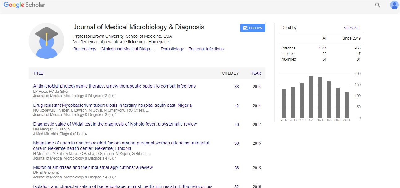
Medical Microbiology & Diagnosis received 14 citations as per Google Scholar report