Opinion - (2022) Volume 7, Issue 2
Received: 01-Mar-2022, Manuscript No. JDCM -22-62901;
Editor assigned: 03-Mar-2022, Pre QC No. P-62901;;
Reviewed: 17-Mar-2022, QC No. Q-62901;;
Revised: 21-Mar-2022, Manuscript No. E-62901;
Published:
29-Mar-2022
, DOI: 10.37421/2475-3211.2022.7.170
Citation: Fatima, Noor. “Angiogenesis in Chronic Wounds in Diabetic Patients.” J Diabetic Complications Med 7 (2022): 170.
Copyright: © 2022 Fatima N. This is an open-access article distributed under the terms of the Creative Commons Attribution License, which permits unrestricted use, distribution, and reproduction in any medium, provided the original author and source are credited.
Neovascularization, commonly characterized as angiogenesis, is vital for wound healing because it involves the formation of granulation tissue by the creation of new capillaries. New capillaries appear in the wound bed as granulation tissue three to five days following tissue injury, acting as a matrix for proliferating blood vessels, migratory fibroblasts, and new collagen. Chronic wounds caused by diabetes and venous or arterial insufficiency are characterised by impaired granulation. Wound healing relies heavily on angiogenesis. Capillary sprouts devour endothelial cells and infiltrate the Extracellular Matrix (ECM) stroma after breaching the underlying Vascular Basement Membrane (VBM), forming tube-like structures that expand, branch, and create networks. Endothelial cell proliferation drives capillary progress in the ECM during angiogenesis, while chemotaxis from the target location guides growth direction. Endothelial cells, angiogenesis factors, and surrounding ECM proteins interact in a time and space-synchronized manner [1,2].
Diabetes patients have aberrant angiogenesis in a variety of organs. Abnormal blood vessel formation (e.g., retinopathy, nephropathy) and accelerated atherosclerosis, which contribute to coronary artery disease, peripheral vascular disease, and cerebrovascular disease, are all vascular diseases linked to diabetes. Angiogenesis is reduced in diabetics, resulting in inadequate development of new blood vessels and, as a result, reduced entry of inflammatory cells and their growth factors. In diabetic wound models, growth hormones including FGF-2 and PDGF, which are important for wound healing, have been reported to be diminished. Furthermore, topical injection of high glucose to wounds in rat models was demonstrated to disrupt the normal angiogenic process, implying that high glucose levels have a direct role in reduced angiogenesis. Wound angiogenesis is a practical model for studying molecular pathways involved in vascular structure creation and remodelling. The repair of a skin defect, in particular, provides a good model for studying angiogenesis since it is simple to regulate and manipulate. Chemical mediators, the ECM, metabolic gradients, and physical forces all operate locally to govern vessel expansion. Modulation of certain parameters is being tried in experimental wounds to promote healing. Scientists are developing mathematical models to understand the involvement of angiogenesis during the healing of (soft tissue) wounds. The macrophage-derived chemical attractant profile, extracellular matrix, and fibroblast diffusion coefficient can all be studied using this model modification of the capillary tip to improve wound healing [3,4].
Venous insufficiency ulcers, also known as venous stasis ulcers, are caused by ineffective valves in the lower leg veins, which cause venous stagnation and hypertension, exposing the skin to ulceration. VEGF stands for vascular endothelial growth factor (VEGF) Patients with chronic venous stasis ulcers have high amounts of VEGF in their blood. This could explain their wounds' enhanced vascular permeability and serum fluid transudation. Biopsies of these ulcers reveal micro capillaries encircled by fibrin cuffs made up of fibrin and plasma proteins such -macroglobulin, which are hypothesised to obstruct gas exchange. Clinical studies have revealed that transcutaneous oxygen tension in venous stasis ulcers can be up to 85% lower than in normal skin regions. Hypoxia increases VEGF expression, which worsens vascular permeability, leads to the creation of pericapillary fibrin cuffs, and compromises gas exchange, reducing growth factor availability in the wound. In granulation tissue, VEGF stimulates the creation of convoluted, aberrant glomeruloid-like vascular structures. Within three days of receiving VEGF, laboratory animals develop these glomeruloid vascular structures, which are characterised by poor perfusion. The persistence of glomeruloid veins in venous ulcers might obstruct oxygen delivery and postpone healing. Proteases such neutrophil elastase, MMPs, and urokinase-type plasminogen activator are abundant in chronic venous stasis ulcers. At the same time, protease inhibitors like plasminogen activator inhibitor-2 are being depleted. Excessive protease activity can damage granulation tissue by degrading growth nutrients [5].
Angiogenesis is a physiological phenomenon which is required for wound healing to occur correctly. Hypoxia, inflammation, and growth factors are all factors that control wound angiogenesis. Angiogenesis' molecular and cellular steps have been identified, and chronic wounds show deficiencies in this process. New wound healing strategies based on this information are emerging to provide growth factors to the wound bed. Surgeons and other wound-care specialists can utilise this information to spot flaws and choose interventions that will help the wound granulate and heal more quickly.
Google Scholar, Crossref, Indexed at
Google Scholar, Crossref, Indexed at
Google Scholar, Crossref, Indexed at
Google Scholar, Crossref, Indexex at
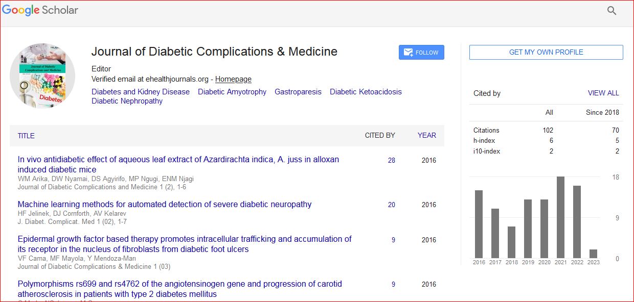
Journal of Diabetic Complications & Medicine received 102 citations as per Google Scholar report