Opinion - (2024) Volume 10, Issue 3
Received: 14-Dec-2022, Manuscript No. JOTR-22-83462;
Editor assigned: 19-Dec-2022, Pre QC No. JOTR-22-83462 (PQ);
Reviewed: 03-Jan-2023, QC No. JOTR-22-83462;
Revised: 24-Mar-2023, Manuscript No. JOTR-22-83462 (R);
Published:
31-Mar-2023
, DOI: 10.37421/2476-2261.2024.10.280
Citation: Bonelli, Zyanya. "Advanced Head and Neck Squamous
Cell Carcinomas that were Previously Untreated and
Might have been Removed are Given Paclitaxel." J Oncol Transl Res 9
(2023): 216.
Copyright: © 2023 Bonelli Z. This is an open-access article distributed under the terms of the creative commons attribution license which permits unrestricted
use, distribution and reproduction in any medium, provided the original author and source are credited.
With surgical resection and postoperative radiotherapy, advanced carcinomas of the oral cavity, oropharynx, and hypopharynx have a 4 years survival rate of 38%, whereas advanced laryngeal carcinomas have a 5 years survival rate of 43%. Although locally advanced head and neck carcinoma is highly responsive to chemotherapy, clinical trials using various induction chemotherapy regimens in patients with operable head and neck carcinoma have failed to demonstrate that the addition of primary chemotherapy results in a significant survival advantage included patients with advanced head and neck carcinoma in their phase I/II trial of increasing the amount of paclitaxel they took every week. Paclitaxel had a starting dose of 40 mg/m2, which was increased by 10 mg/m2/week up to a maximum of 90 mg/m2. There was one patient with a Complete Response (CR), two Partial Responses (PRs), four with Stable Disease (SD), and one with Progressive Disease (PD) among the eight patients with head and neck carcinoma. The Eastern Cooperative Oncology Group (ECOG) began a phase II trial of paclitaxel 250 mg/m2/24 hours infusion every 21 days with colony stimulating factor primary prophylaxis in 34 patients with recurrent, metastatic, or locally advanced, incurable squamous cell carcinomas of the head and neck. After registration was completed, four patients were deemed ineligible. 28 of the 30 eligible patients were examined for response, having received an average of four courses of chemotherapy: 8 PRs, 9 SDs, 7 PDs, and 4 CRs all eligible patients had a median response time of 4.5 months, while patients with a CR had a median response time of 12 months [1].
The 1 year survival rate was reported to be 33% (90% CI, 19– 47%), and the median survival rate for all eligible patients was 9.2 months. Thirty-three of the 33 patients who could be examined for toxicity had severe or life-threatening granulocytopenia, and three of them had an infection that was severe or life-threatening. Three, four, and one patient each experienced severe stomatitis, neurosensory toxicity, or neuromotor toxicity, respectively. There was one death from typhlitis, one from septicemia, and one from myocardial infarction.
Paclitaxel was found to be one of the most effective single agents for the treatment of head and neck carcinoma in this study, which was conducted on patients whose cancer was far advanced and had been treated previously [2].
Cisplatin containing induction chemotherapy regimens were used most frequently in head and neck carcinoma patients. We proposed investigating paclitaxel as induction therapy in the advanced resectable head and neck carcinoma patient population due to its promise in the ECOG trial in patients with metastatic, recurrent, or locally advanced incurable squamous cell carcinomas of the head and neck. As a result, the primary objective of this study was to ascertain the rate of response to paclitaxel as a single-agent induction therapy and the amount of time it took for progression. We discuss the function of paclitaxel in this particular clinical setting and discuss the outcomes of the 45 participants in this trial [3].
Patients were ineligible if they had a history of cancer within the previous five years (with the exception of basal or squamous carcinomas of the skin and carcinoma in situ of the cervix that had been adequately treated), significant cardiac disease (such as angina pectoris, congestive heart failure, cardiac arrhythmias, second or third degree heart block, bundle branch blocks, or myocardial infarction within the preceding six months), current use of any medication that could affect A multimodality team that included a head and neck surgeon, a medical oncologist, and a radiation oncologist examined all of the patients. Prior to any therapeutic intervention, the patient's head and neck surgeon was required to plan the resection based on the extent of the cancer [4].
Before beginning treatment, written informed consent was obtained from each patient. When an adverse event occurred that, in the opinion of the investigator, prevented further participation in the protocol, patients were removed from the study, or when the patient requested it. The trial's design called for complete surgical resection of patients four weeks after the last dose of paclitaxel was given. The extent of the surgical resection was determined using the initial prechemotherapy tumor extent. The affected region determined the distance between the surgical margins and the primary tumor. A pathologist examined the surgical margins of each primary tumor bed during the procedure. The patient provided the tissue samples, not the specimens, and the frozen section technique was utilized for slide preparation. Most of the patients had at least one radical ipsilateral neck dissection. In the event that the carotid adventitia was to be cut out, dermal grafts were used to shield the carotid artery. Primary closure of the surgical defect was used whenever possible. Musculocutaneous flaps or osteocutaneous free flaps were used to reconstruct surgical defects during extensive resections. Plates for mandibular stabilization were also used. Nonaminoglycoside intravenous antibiotics were administered prior to, during, and for 48 hours after the procedure [5].
After the patients recovered from their surgeries, they would begin receiving external beam radiotherapy. All patients received megavoltage radiotherapy of greater than 1 MV. Cobalt-60 was used to treat three patients, and a linear accelerator was used to treat the remaining ones, using 4-8 MV X-rays for the primary/neck fields and electrons for the neck boosts. Custom cerrobend blocks were used as additional shielding, and the patients were simulated prior to treatment beginning. All fields were treated every day (Monday through Friday) at a distance of less than 80 cm. Conventional fractionation consisted of once daily treatments using 1.8-2 gray (Gy) fractions. The upper neck draining lymphatics and the site of the previous primary tumor were treated with a parallel opposed or wedged pair technique. A single AP field was used to treat the lower neck and supraclavicular areas, and it was set to D-Max or 3 cm depth. The dose to the prior primary tumor site ranged from 45 to 70.2 Gy, with a median of 55.8 Gy, and the dose to the lymph node regions from 45 to 70 Gy, with a median of 50.4 Gy. The total dose to the spinal cord could not exceed 45 Gy.
[Crossref] [Google Scholar] [PubMed]
[Crosssref] [Google Scholar] [PubMed]
[Crossref] [Google Scholar] [PubMed]
[Crossref] [Google Scholar] [PubMed]
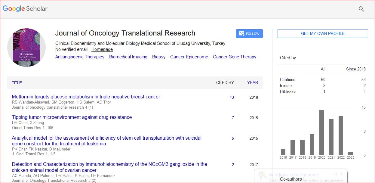
Journal of Oncology Translational Research received 93 citations as per Google Scholar report