Commentary - (2022) Volume 11, Issue 2
Received: 05-Feb-2022, Manuscript No. jio-22-55102;
Editor assigned: 07-Feb-2022, Pre QC No. P-55102;
Reviewed: 11-Feb-2022, QC No. Q-55102;
Revised: 17-Feb-2022, Manuscript No. R-55102;
Published:
28-Feb-2022
, DOI: 10.37421/2329-6771.2022.11.364
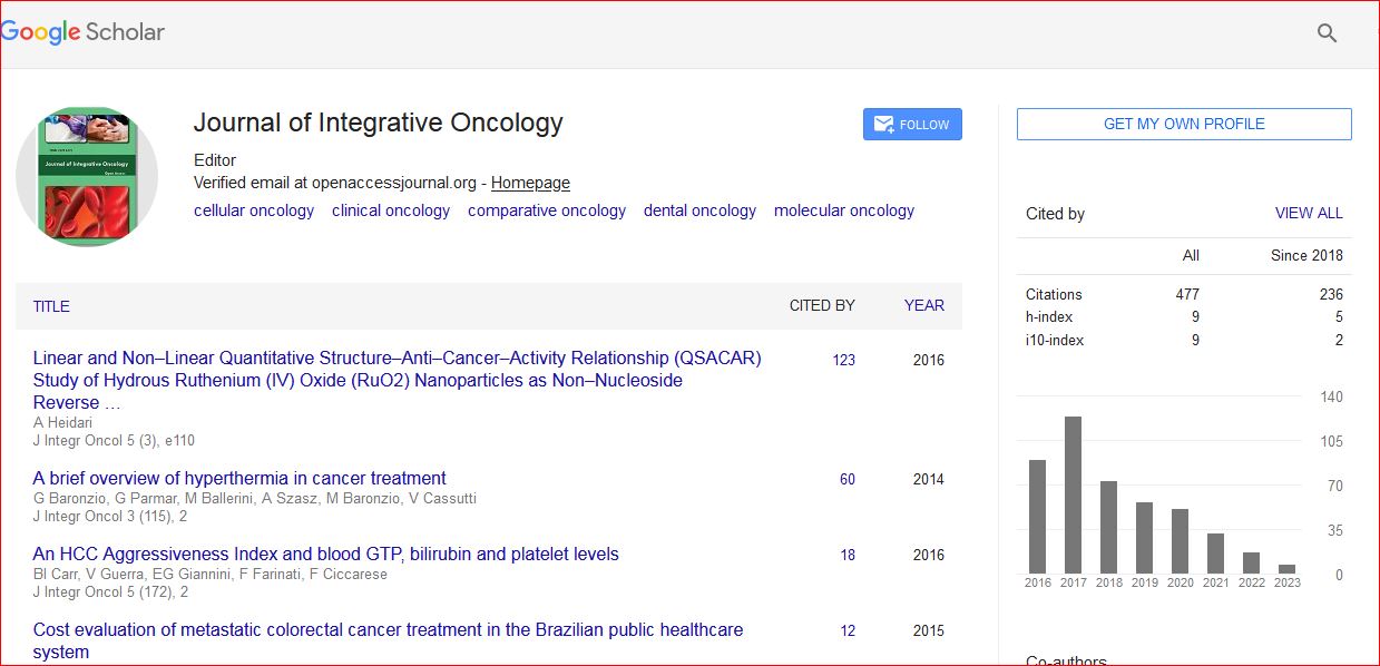
Citation: Ren, Jiang. “About Cancer-associated Fibroblasts (CAFs) and Cancer Markers.” J Integr Oncol 11 (2022): 364. DOI: 10.37421/2329-6771.2022.11.364
Copyright: © 2022 Ren J. This is an open-access article distributed under the terms of the Creative Commons Attribution License, which permits unrestricted use, distribution, and reproduction in any medium, provided the original author and source are credited.
The majority of the tumour bulk in pancreatic ductal adenocarcinomas is made up of cancer-associated fibroblasts (CAFs) (PDACs). The majority of current efforts to eliminate malignant tumours target the proliferation of fast developing cancer epithelial cells. We know that this is largely useless, as most cancers develop resistance to treatment after exposure. Despite the fact that CAFs have long been recognised as important in PDAC, little is known about how chemotherapy affects CAFs and how they may contribute to drug resistance in adjacent cancer cells. We show here that CAFs exposed to chemotherapy play an important role in cancer cell survival and proliferation. CAFs are innately resistant to gemcitabine, the chemotherapeutic standard of therapy for PDAC, according to our findings.
Furthermore, CAFs treated to gemcitabine produce more exosomes, which are extracellular vesicles. In recipient epithelial cells, these exosomes enhanced the chemoresistance-inducing factor Snail, which promotes proliferation and drug resistance. Finally, treating gemcitabine-exposed CAFs with GW4869, an inhibitor of exosome release, lowers survival in co-cultured epithelial cells, indicating that CAF exosomes play a key role in chemotherapeutic drug resistance. Overall, our findings suggest that exosome inhibitors could be used in conjunction with chemotherapy to overcome PDAC chemoresistance. Despite the presence of non-neoplastic stromal components that significantly contribute to tumour growth, efforts to create anti-cancer medicines have mostly concentrated on targeting the epithelial compartment. The surrounding tumour microenvironment has an impact on cancer cell survival, proliferation, migration, and even dormancy (TME). Cancer-associated fibroblasts (CAFs) have been found to have a number of roles in the formation of tumours within the TME.
Growth factors, inflammatory ligands, and extracellular matrix proteins are all secreted by cancer cells, promoting cancer cell proliferation, therapeutic resistance, and immune exclusion. However, new research suggests that CAFs may be able to slow tumour development in some cases. We summarise the corpus of knowledge on CAFs in this review, with a focus on the most recent discoveries about fibroblast heterogeneity, plasticity, and roles. We also emphasise the similarities among fibroblasts found in various cancer types, as well as in normal and inflammatory states. Finally, we discuss the most recent developments in preclinical and clinical trials of therapeutic techniques targeting CAFs. One of the main elements of tumour development and invasion has been identified as the tumour microenvironment.
Cancer-associated fibroblasts (CAFs), a type of perennially activated fibroblast, have been implicated in this milieu for having a significant tumormodulating effect and playing a key role in areas like treatment resistance. The expression of different "CAF indicators," such as Fibroblast Activation Protein Alpha (FAP) and Alpha Smooth Muscle Actin (SMA), has traditionally been used to distinguish CAFs from the greater pool of fibroblasts found in the body. The expression of numerous commonly used fibroblast markers, on the other hand, is exceedingly diverse and varies significantly between CAF subpopulations. As a result, novel selection strategies based on cellular function, as well as future study characterization, are critical for standardising CAF identification and improving the cross-applicability of diverse research investigations in the field. The goal of this review is to provide a comprehensive overview of the most regularly utilised fibroblast markers in the field, as well as their varied strengths and drawbacks, as well as prospective future pathways for CAF identification and targeting.
Long-term investigations of cancer-associated fibroblasts (CAFs) and their interactions with cancer stem cells, one of the primary driving forces in patients with oral squamous cell carcinoma, have been hampered by the limitations of the current model (OSCC). The 3-dimensional organoid model was used in this work to see if co-culturing with paralleled CAFs enhances stem-like characteristics in OSCC. Patients with OSCC were used to create tumour organoids and corresponding CAFs in the lab. The CD44+ cells in organoids were then flow cytometrically separated and co-cultured with CAFs in Matrigel. The ability of CD44+ cells to form organoids is enhanced when they are co-cultured with CAFs, and this effect is reduced when lactate generation or absorption in CAFs or CD44+ cells is suppressed. In addition, CD44 and OCT-4 expression levels in organoids incubated with lactate were measured using immunofluorescence or a Western blot analysis. The findings showed that lactate treatments improve CD44+ cells' ability to produce organoids as well as the protein expression of CD44 and OCT-4 in OSCC organoids [1-5].
Google Scholar, Crossref, Indexed at
Google Scholar, Crossref, Indexed at
Google Scholar, Crossref, Indexed at
Journal of Integrative Oncology received 495 citations as per Google Scholar report