Mini Review - (2022) Volume 13, Issue 11
Received: 03-Nov-2022, Manuscript No. jhmi-23-86859;
Editor assigned: 05-Nov-2022, Pre QC No. P-86859;
Reviewed: 16-Nov-2022, QC No. Q-86859;
Revised: 22-Nov-2022, Manuscript No. R-86859;
Published:
30-Nov-2022
, DOI: 10.37421/2157-7420.2022.13.447
Citation: Estrom, Antte. “A Mini Review on Application for describing Dental Conditions.” J Health Med Informat 13 (2022): 447.
Copyright: © 2022 Estrom A. This is an open-access article distributed under the terms of the Creative Commons Attribution License, which permits unrestricted use, distribution, and reproduction in any medium, provided the original author and source are credited.
Healthcare could significantly benefit from improved communication; It is possible that patients who are better informed are more likely to participate in the management of their health conditions, make better decisions based on their knowledge, and ultimately contribute to improved quality of care. The use of pictures enhances knowledge, understanding, and recall, according to a recent systematic review and meta-analysis of the effects of using visual aids to convey health information on patient and consumer health behaviors and outcomes. Numerous clinical trials have demonstrated the efficacy of visual aid-based interventions designed to enhance patient comprehension and education regarding surgical procedures and the management of chronic diseases.
Health care • Dental condition • Density
This is especially true for individuals who are low in literacy and for situations in which visualization is crucial. New technologies offer additional opportunities for enhanced visual communication based on these modalities. In clinical and surgical settings, web-based and multimedia-augmented patient education tools were evaluated in positive randomized controlled trials. Communication tools like these are said to be better than printed, written information, like pamphlets, and they may help patients feel better about giving informed consent and other aspects of a clinical encounter. In dentistry and oral health care as a whole, patient education is of the utmost significance. Education and communication are fundamental to promoting oral health because of the nature of common dental diseases like dental caries and periodontal disease, which have strong behavioral and selfcare components and are chronic but preventable. In essence, Albano and colleagues argue that patients should be able to improve their health behaviors and self-care strategies if they have a better understanding of their illness and treatment, which may even reduce treatment-related costs. When education is targeted at parents and relates to their children's diseases and surgical treatment, the potential may be even greater. The advantages of visual communication aids as tools for enhancing parents' and children's understanding of dental conditions and associated treatments have recently been demonstrated in pediatric dentistry. In a recent study, both video and physical models were found to improve hearing-impaired children's oral hygiene.
Sculptor, a web-based data visualization informatics pipeline that aims to improve visual communication in dentistry and oral health care for children, is presented in this communication along with its development, deployment, and two real-world applications. A three-dimensional, lifelike model of the pediatric dentition that can be dynamically annotated to illustrate dental conditions (such as "cavities" or missing teeth) and existing or proposed restorative treatments (such as fillings and crowns) was the primary objective of our efforts. The 3D model is expected to be useful in clinical, research, and dental education settings for one-on-one education with families, illustrating clinical findings and proposed treatment plans, and making the informed consent process easier. In addition, we anticipate that it can be modified to function as an effective visual tool for summarizing the outcomes of epidemiological surveys or other dental research studies that produce information at the surface level of teeth. We talk about the performance, public deployment, design and customization features, and two real-world applications of the pipeline in the following sections. The twenty primary human teeth are included in our model; these are divided into 88 distinct tooth surfaces in accordance with dental anatomy principles, treatment planning conventions, electronic patient records, and documentation requirements.
A pipeline that can quickly apply a subset of modifications to an existing 3D model is described in the following paragraphs. The modifications, which are specified in advance and unique to the fundamental model, may include, but are not limited to changes in color, regional opacity, and how parts of the model reflect light are all factors. The most recent modification can be used to represent a variety of materials, including enamel a healthy tooth surface metal, glass, porcelain, and so on. Using a key that maps input data to modifications; modifications can be applied automatically once they have been defined. The initial model is derived into the output. Understanding the general structure of 3D models and, more specifically, the structure of the model to be modified is the first step in building the modification interface.
Importing and creating models from individual tooth surface-level data of 54 ECC subtypes (similar to those shown in Figure 5) was then used to evaluate the prototype's speed. 54 annotated 3D renditions of the primary dentition were generated and displayed by the pipeline at an average rate of 956 milliseconds per model, with a standard deviation of 58 milliseconds. Compared to the alternative strategy of manually editing the base model, which necessitates not only specialized knowledge of the model but also orders of magnitude more time, this amount of time is negligible. Based on these two real-world applications, we hypothesize that SculptorHD's methods and web-based deployment are ideal for creating lifelike 3D models of the pediatric dentition that can be used for a variety of downstream applications, such as clinical, patient education, and research, while also allowing for additional customization and post-processing of the produced models [1-6].
Models have a long history of helping us understand the world. In this paper, we present a customizable method for creating better 3D models that can be immediately used in pediatric dentistry and oral health. In clinical, educational, and research settings, we strongly believe that the SculptorHD model will be practically useful as a communication tool for visualizing tooth-surface level conditions of the pediatric dentition. There are currently no empirical data to determine its utility, acceptability, or superiority over more established methods like printed material, other than the two real-world applications that we presented. We find that the tool is sufficiently quick and adaptable to be used as a chair side communication aid and interface with an electronic patient record, in addition to its presumed attractiveness for presenting clinical findings and research results. It is also possible that educational modules will incorporate it.
We must emphasize that the tool is not meant to replace personalized, direct intraoral 3D scans of each patient. Clearly, the latter would be superior in terms of being entirely personalized and illustrating dental conditions with greater specificity. However, 3D scanning for the sole purpose of documentation is not yet performed on a regular basis in clinical, public health, or research settings and it may be particularly challenging for very young children. Despite this, we believe that the fact that our model relies on conventionally obtained, tooth surface-level data on dental caries, as well as actual or planned restorations, may make it possible to facilitate interactions between patients and providers. In addition, the instrument is adaptable enough to depict a wide range of conditions, including hypoplastic defects, non-carious tooth surface defects, findings from radiographic examinations, such as proximal caries lesions, and other potential pathologies. We strongly believe that this strategy has significant potential to enhance the overall quality of dental care delivery and that we have demonstrated its value in the real world through two applications. In conclusion, we were able to successfully design, develop, and web-deploy a 3D visualization tool with customizable user inputs and outputs for illustrating the surface conditions of pediatric teeth in life-like models of the pediatric dentition.
None.
The authors declare that there is no conflict of interest associated with this manuscript.
Google Scholar, Crossref, Indexed at
Google Scholar, Crossref, Indexed at
Google Scholar, Crossref, Indexed at
Google Scholar, Crossref, Indexed at
Google Scholar, Crossref, Indexed at
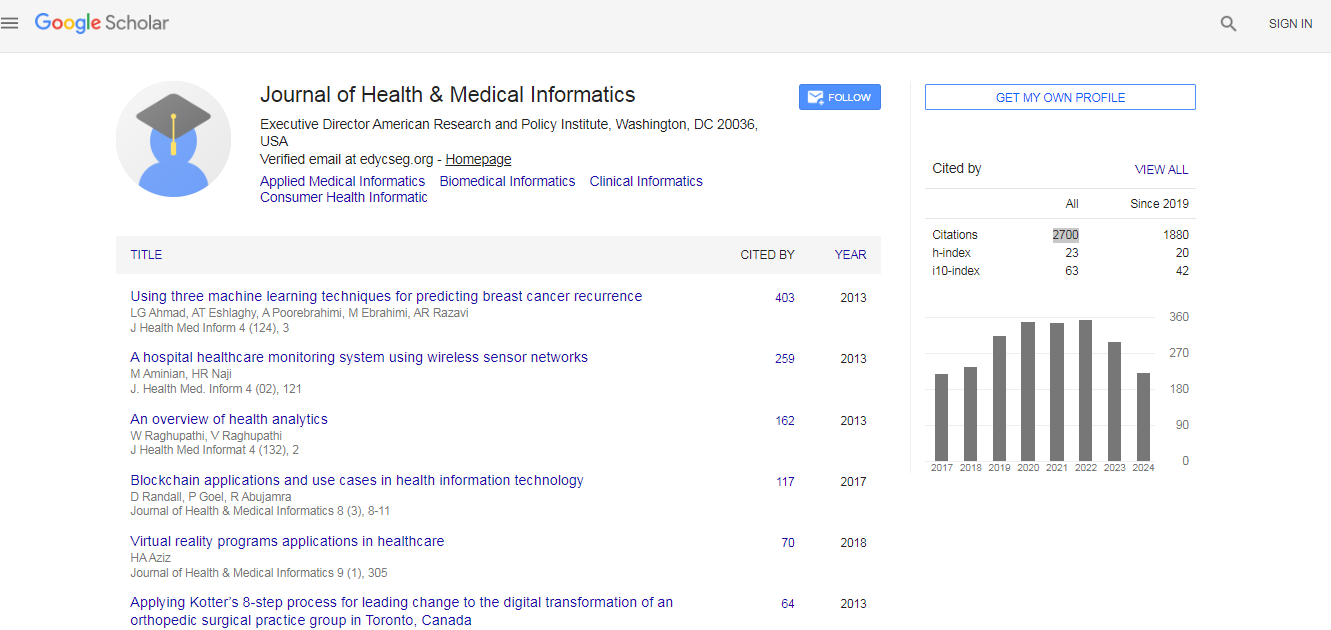
Journal of Health & Medical Informatics received 2700 citations as per Google Scholar report