Gary Greenberg
University of Hawaii Institute for Astronomy, USA
Keynote: J Laser Opt Photonics
When Marvin Minsky invented confocal microscopy in the 1950�??s, he likely did not envision the huge impact it would have on biomedical research in the 21st century. Confocal technologies have been essential for our modern understanding of how DNA, proteins, enzymes and cells function in health and disease. Confocal technologies have overcome the greatest problem with conventional microscopes, which is their extremely shallow depth of field. A high-power light microscope has a depth of field of only a few microns or less. Traditionally, microscopists cut tissue samples into 5 micron sections for the purpose of reducing blur from out-of-focus structures. This conventional approach of examining 5 micron thick sections introduces a significant sampling error when looking at biological tissue because a single cell is about 20 microns in diameter. The results produce incomplete images that reveal only a small portion of a single cell. The great benefit of confocal microscopes is their ability to dramatically increase the depth of field though stacking images from different focus levels and then reconstructing the stack of pictures into 3D images with improved sharpness and removal of out-of-focus blur. These three-dimensional microscopes provide a clear view of a thick volume of tissue, up to 100 microns thick or more. The volume of thick tissue can be observed from multiple points of view, providing significantly more information about the specimen being examined. The result is increased productivity, better diagnoses and improved understanding. Edge-3D is a non-confocal light microscope that provides most of the advantages of expensive confocal instruments at a fraction of the cost and complexity of operation. Out-of-focus blur is removed using software algorithms that are compatible with a range of optical systems, including, reflected illumination, transmitted illumination, oblique illumination, brightfield, darkfield, phase contrast, DIC, polarization and livecell imaging. Multiple modes of 3D display, including real-time 3D imaging, expose hidden depth information and reveal the relationships between different structures within the specimen being observed. An additional benefit is the ability to measure the structures within the specimen in 3D. He will talk about the history of 3D microscopy and present dramatic 3D images from a range of application areas, including, neurobiology, plant biology, entomology, forensic sciences and the geology of the lunar sand.
Gary Greenberg earned a PhD in Developmental Biology from University College London in 1981. He was an Assistant Professor at the University of Southern California during the 1980’s. In the early 1990’s he began inventing and developing three-dimensional light microscopes, for which he holds 20 US patents. He is the CEO of Edge-3D, LLC, a company that innovates 3D imaging devices for research, industry and biomedicine. He has published numerous peer-reviewed journal articles and has given over fifty international talks, including two TED talks. He has written two books about sand through the microscope, A Grain of Sand: Nature’s Secret Wonder (2008) and The Secrets of Sand (2015) and a children's book, Mary's Magic Microscope (2011). He is currently a Research Affiliate at the Advanced Technology Research Center, the University of Hawaii Institute for Astronomy in Maui, where he uses 21st-century microscopes to study Moon sand brought to Earth by NASA during the Apollo Missions. Each grain of sand is unique and each has a story to tell.
E-mail: Greenberg@edge-3D.com
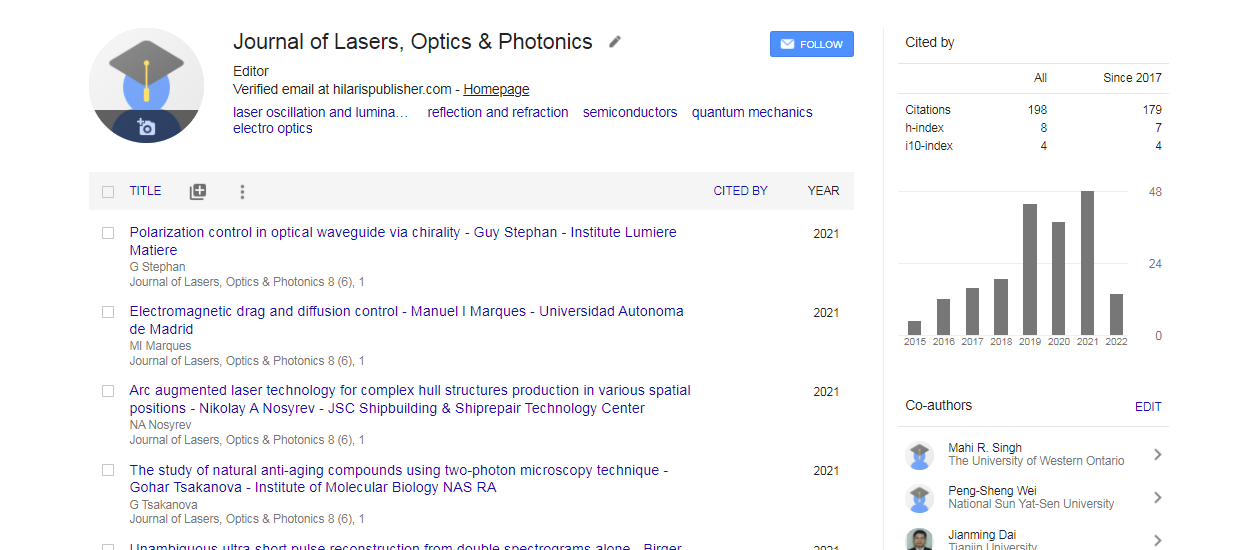
Journal of Lasers, Optics & Photonics received 279 citations as per Google Scholar report

 Spanish
  Chinese
  Russian
  German
  French
  Japanese
  Portuguese
  Hindi
Hindi 