Giuseppe Antonacci
Centre for Life Nano Science, Italy
Scientific Tracks Abstracts: J Laser Opt Photonics
Brillouin spectroscopy has shown great potential to become a reliable diagnostic tool due to its capability of measuring viscoelastic properties of materials in a non-contact manner. The recent development of high-sensitivity CCD cameras and Virtually Imaged Phase Array (VIPA) etalons has dramatically reduced data acquisition time to ~0.1 sec per spectrum. This has brought Brillouin spectroscopy from a point sampling technique to a new imaging modality. We describe the characterization of a confocal Brillouin microscope designed to measure mechanical properties of biological tissues. The frequency broadening of the Brillouin spectrum due to high illumination and collection apertures has been investigated in order to determine the optimal geometry that maximizes both the spectral and the optical resolution. A high extinction ratio was achieved in a Michelson interferometer to suppress strong specular reflections. Sub-micron resolution Brillouin images of single cells and arterial wall tissues have been acquired, in particular when atherosclerotic plaques were formed. These results might encourage the application of Brillouin microscopy as a tool of choice in clinical practice.
Giuseppe Antonacci has completed his PhD from Imperial College London. He is a Research Associate at the Italian Institute of Technology and is an Editor for the De Gruyter Physics.
Email: giuseppe.antonacci@iit.it
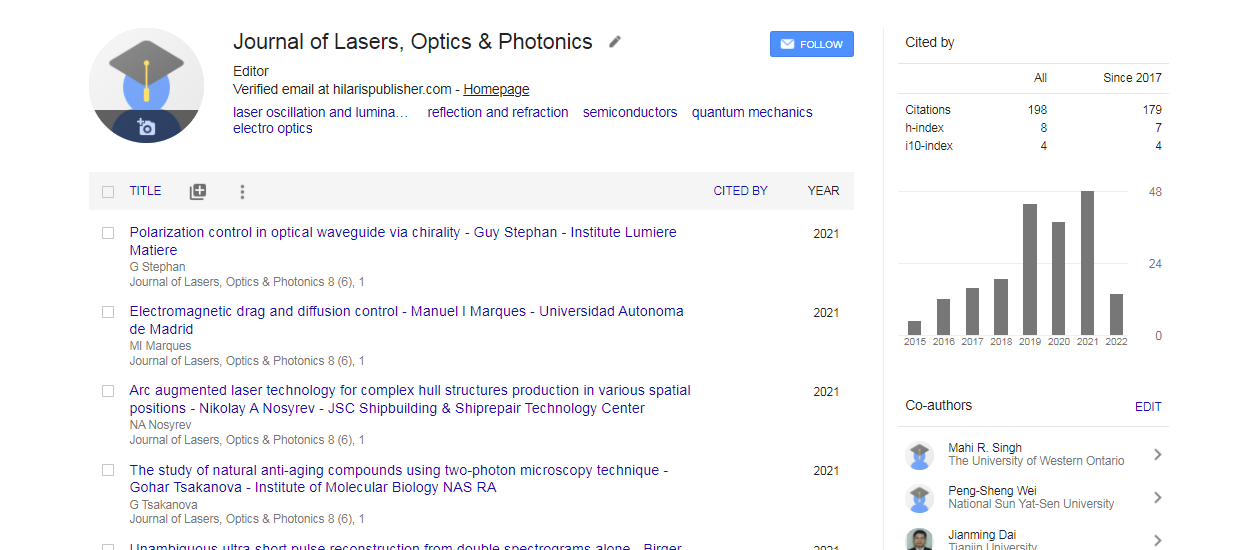
Journal of Lasers, Optics & Photonics received 279 citations as per Google Scholar report

 Spanish
  Chinese
  Russian
  German
  French
  Japanese
  Portuguese
  Hindi
Hindi 