Opinion - (2023) Volume 10, Issue 2
Received: 03-Jan-2023, Manuscript No. ijn-23-88509;
Editor assigned: 04-Jan-2023, Pre QC No. P-88509;
Reviewed: 16-Jan-2023, QC No. Q-88509;
Revised: 21-Jan-2023, Manuscript No. R-88509;
Published:
30-Jan-2023
, DOI: 10.37421/2376-0281.2023.10.506
Citation: Kim, Jeansok. "An Extensive Overview of the Fundamental Concepts and Clinical Implications of Stress Neurobiology." Int J Neurorehabilitation Eng 10 (2023): 506.
Copyright: © 2023 Kim J. This is an open-access article distributed under the terms of the Creative Commons Attribution License, which permits unrestricted use, distribution, and reproduction in any medium, provided the original author and source are credited.
Controlling thoughtful outpourings that are involved in enhancing response to external pressure and directing glucose homeostasis are controlled by mind administrative components. A few crucial brain runs have been defined in terms of the fundamental subatomic systems, downstream neurocircuitries, and the issues at hand through research in this rapidly developing field. It has been hypothesized that persistent pressure, such as openness to erratic stressors or ongoing social loss stress, alters synaptic designs in the prefrontal cortex and the basolateral amygdala (BLA). The ability of the hypothalamic-pituitary-adrenal hub predicts that changes in the hypothalamus will occur in response to high or persistent strain. Other thought runs, such as the basolateral amygdala, which contributes to stressinduced psychopathology, have been described by subsequent research. Using the extreme limit pressure model, however, there are no planned pressure networks in the entire brain to separate the central role of the important cerebrum rons in pressure and transformation. Using c-fos as a journalist, we profile important mind cores involved in the CNS response to strong pressure in this paper. These mind cores may provide unique focuses on the issue of intense pressure [1].
The chemicals that are delivered pressure have an effect on how glucose is broken down. After being put under a lot of pressure, cortisol levels usually went up. After pressure openness, blood glucose and insulin levels went up, which suggested that weight has a negative effect on blood glucose homeostasis. We show that the brain responds differently to high glucose levels and high limit pressure in this study, indicating that the brain is primarily responsible for glucose homeostasis rather than a second response to push [2].
Another type of physiological pressure actuates endoplasmic reticulum stress, acting through anticipation of N-glycosylation of proteins, and may direct cell reactions to oxidative pressure and cause cytotoxicity. A nonmetabolizable type of glucose enters the cell and represses glycolysis. It is used to mimic hypoglycemia. A robotic connection between hypoglycemia and development chemical delivery, a type of pressure reaction, was suggested after it was demonstrated that hypoglycemia enacts GHRH neurons in the cerebrum runs, such as the periventricular core. The majority of the research conducted by scientists in these works focused on the primary components of glucose detection and the guidelines for glucose metabolic homeostasis. To identify the Ronal controls in the cerebrum under these two distinct physiological loads, we profile and consider mind maps triggered by intense limit pressure and mixture in this section [3].
First, we examine the pressure-controlling CNS cores in the brain. Laine et al., for instance, demonstrated explicit mind runs associated with various pressure conditions in previous research. have shown that constant social loss causes changes in certain parts of the mind. One of the best things about our review is that it uses cutting-edge imaging and examination techniques to focus on preoperative brain volumes, perfusion, and infarcts according to postoperative wooziness. This is the primary focus of the investigation into the ridiculousness and shape of WMH. Because they were not approved for use in between-focus applications, these WMH shape markers were not examined in the entire review group. Additionally, our review included a diverse group of patients scheduled for a variety of major medical procedures from two review environments, which increased the generalizability of our findings [4].
The animal care and use guidelines established by the Shenzhen Institute of Advanced Technology, Chinese Academy of Sciences, were followed in all of the analyses of the creatures. We used self-control pressure, a conventional pressure paradigm, to energize mice following the restriction stress excitement in order to distinguish the specific cerebrum regions that responded to the outer physiological rather than the actual pressure; In pushed mice, we examined the articulation throughout the entire cerebellum. Our c-fos staining was effective because a significant number of fluorescence cells were found in a few mind regions. We thought about the covered or special runs response to pressure answering, 2DG- and glucose-detecting in the CNS by C-fos marking at the entire mind map book separately in the current review [3].
The broad focus on convention for all members, which may have presented a selection of patients who were less defenseless in comparison to patients who declined cooperation, may be one of the review's limitations. The observed correlation between preoperative MRI highlights and postoperative wooziness may have been misinterpreted by this. We may have to exclude patients with head movement concerns, particularly for the perfusion MRI, as an additional restriction. This made it harder to differentiate between bunch contrasts, which may have led to the rejection of weak patients who were unable to remain still in the MRI scanner. In any case, there were no differences in the frequency of drowsiness between the group that underwent our perfusion examination and the group that was rejected [5].
The fact that not all sweeps could have been used for some of the cerebrum MRI results may have undervalued the found results for some of these highlights. Another limitation could be that we used two distinct types of MRI scanners, which resulted in an expected contrast between the focus areas. Nevertheless, in all of our investigations, we utilized a picture examination pipeline that is well-suited for focus contrasts. Unfortunately, we were unable to determine whether members' cortical infarcts were indicative of their injuries. In addition, we lacked the measurable capability to conduct examinations of the impact of injury area due to the large variety of sore areas and the small number of patients with cortical infarcts.
None.
None.
Google Scholar, Crossref, Indexed at
Google Scholar, Crossref, Indexed at
Google Scholar, Crossref, Indexed at
Google Scholar, Crossref, Indexed at
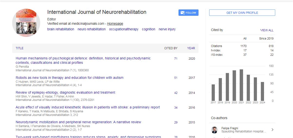
International Journal of Neurorehabilitation received 1078 citations as per Google Scholar report