Chulkova Svetlana Vasilievna1,2
Most of the cancers are still incurable human diseases. All malignant tumors are a heterogeneous population of cells with different biological properties. There are two dominant concepts to explain tumor heterogeneity: the theory of tumor stem cells (TSCs), also called the hierarchical model and the stochastic model. TSCs constitute only a small percentage (0.05-1%) of tumor cells within a tumor mass containing heterogeneous population of tumor cells within the tumor microenvironment. TSCs are closely related to pathological features which result in worse clinical prognosis. TSCs are distinguished by the pronounced expression of anti-apoptotic proteins and the high activity of chemoresistance mediators – ABC-1,2,5, desregulation of signaling pathways Notch, Hedgehog, Wnt. TSCs harbour endogenous resistance mechanisms against radiation and chemotherapy which gives TSCs a survival advantage over differentiated counterparts. The currently known 40 TSC surface markers can express on the hESCs, adult stem cells, and normal tissue cells. Of the 40 TSC markers, approximately 83% (33 out of 40 TSC markers) are rarely expressed on normal tissue cells. 9 of these are already approved as drug target molecules by FDA. A minor TSC subset with maximum resistance to conventional anticancer therapies plays a special role in metastasis. According to the literature, melanoma TSCs are characterized by expression of antigens such as CD44, CD271, and CD133. CD133 is the most frequently studied TSC surface marker in various cancers. In our study we identified TSCs among melanoma DTCs by CD133 expression. 47 bone marrow samples of patients with melanoma were analyzed before treatment by flow cytometry. Among them, stage IV was established in 42.6% of cases. The immunophenotype of the cells was characterized based on the expression of the following antigens: Syto41, CD45, HMB-45, CD133. Evaluation of the expression of antigens was performed within the nucleated cells of Syto41+. Among the evaluated bone marrow samples, the presence of Syto41+CD45-HMB-45+ cells was found in 57.4% of cases. Only in one case, the presence of Syto41+CD45- HMB-45+CD133+ cells was established. The percentage of this subpopulation in this sample was 1.28. It should be mentioned in most cases, patients had an advanced stage of melanoma. This may confirm that CSC marker-negative or marker-positive cancer cells could initiate tumor formation. As demonstrated in our study, flow cytometry with a specific antibody HMB-45 in combination with CD45 is a useful technique to assess BM involvement in melanoma. BM involvement was found in 57.4% of skin melanoma cases. In 28.6% of cases I stage disease was established, which confirms the aggressiveness of skin melanoma even in localized disease. BM as a niche for micrometastasis plays a key role in hematogeneous dissemination. The detection of early dissemination of the tumor process may be a step towards individualization of treatment in this category of patients. At the same time, the identification of cells disseminated in the bone marrow with subsequent testing for the presence of TSCs antigens opens up prospects for the development of methods for influencing melanoma on the TSCs. It is nown that diversity of TSCs may be generated by distinct stemness or reprogramming signalling activations, resulting in divergent expression patterns or TSC markers. Recent findings demonstrates that the TSCs can be newly generated from the differentiated non-TSCs by reprogramming mechanism: even TSCs with different characteristics could emerge. TSCs not only serve as the origin of tumor formation but also drive heterogeneity of cell composition inside the tumor and TSCs themselves. In our study the findings of CD57 and CD133 expression are evidence of TSCs heterogeneity and the complex hierarchical relations between the primary tumor and the dissiminated TCs.
PDFShare this article
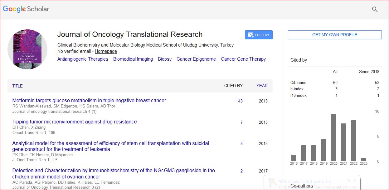
Journal of Oncology Translational Research received 93 citations as per Google Scholar report