Meena Bedi, Jordan Kharofa, Eduardo V Zambrano, Jason Chang, Keith Baynes, Alan P Mautz, Melissa DuBois, David M King, Donald A Hackbarth and Dian Wang
Purpose: MRI is often used to evaluate sarcoma response to neoadjuvant treatment, however its role to predict for pathologic response and survival is unclear.
Methods and materials: From 2003-2010, 116 patients with STS were treated with neoadjuvant therapy (NAT). 62 patients who had an MRI before and after radiotherapy were analyzed. Radiographic change was correlated with survival and necrosis and fibrosis on pathology. ROC curve analysis was used to assess change in volume that best predicted for pathological necrosis.
Results: Median follow-up was 33 months. There was median tumor volume decrease of 15.08 cm3 after treatment. Increase in tumor size and volume was associated with greater necrosis (p<0.03, p=0.001, respectively) and less fibrosis (p<0.001) on pathology. High-grade tumors had more necrosis (p<0.001) and comprised the majority of patients with tumor increases following NAT (88%). Tumor increase of at least 66% predicted for ≥ 70% necrosis with 94% specificity. The 3-year OS was 65% vs. 93% in patients with a decrease in size and volume (p=0.004). In tumors with ≥ 70% necrosis, the 3-year OS was 38% vs. 91% if necrosis was <70% (p<0.001).
Conclusions: MR-based tumor increase following NAT was associated with greater % necrosis and less fibrosis on pathology. This tumor increase was more likely high-grade and associated with worse survival.
Share this article
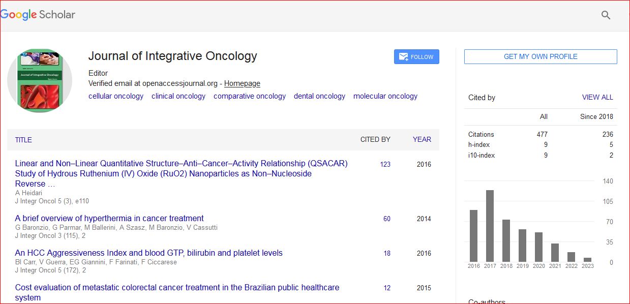
Journal of Integrative Oncology received 495 citations as per Google Scholar report