Rani Kanthan
Pathological analysis and evaluation of a liver biopsy is a crucial step within the diagnosis of single or multiple mass lesions within the liver. Accurate diagnosis is paramount in guiding appropriate treatment. This study conducted an enquiry for liver biopsies for the past 6 years with the diagnostic search codes of neoplasm, metastases, metastatic, adenocarcinoma, neuroendocrine carcinoma, sarcoma, and lymphoma. The aim was to review their pathological workup with a view to developing cost-efficient immunohistochemical diagnostic algorithms. a complete of 375 consecutive neoplastic liver biopsies were retrieved and subjected to pathological review. for sure the bulk up to 95% of the neoplastic lesions were metastatic lesions. some biopsies up to fifteen represented primary hepatocellular
/cholangiocarcinoma, haemangioma, and cirrhosis. the most typical metastases [upto 61%] to the liver were colorectal in origin being Hepar-ve, CDX2+ve, and CK20+/CK7-ve. Other lesions included metastases from pancreas [12%], lung [8%] upper gastrointestinal [8%], neuroendocrine lesions [8%],
ovarian [1%] and kidney/urothelial [2%]. Uncommon metastases encountered included hepatic metastatic meningioma, endometrial stromal sarcoma, and osteosarcoma. Immunohistochemical stains were the foremost useful test in identifying the first site of the tumor. Though diagnostic algorithms were developed especially within the case of the unknown primary, some biopsies received a medical diagnosis of over one organ because the primary site for clinicopathological correlation.
Share this article
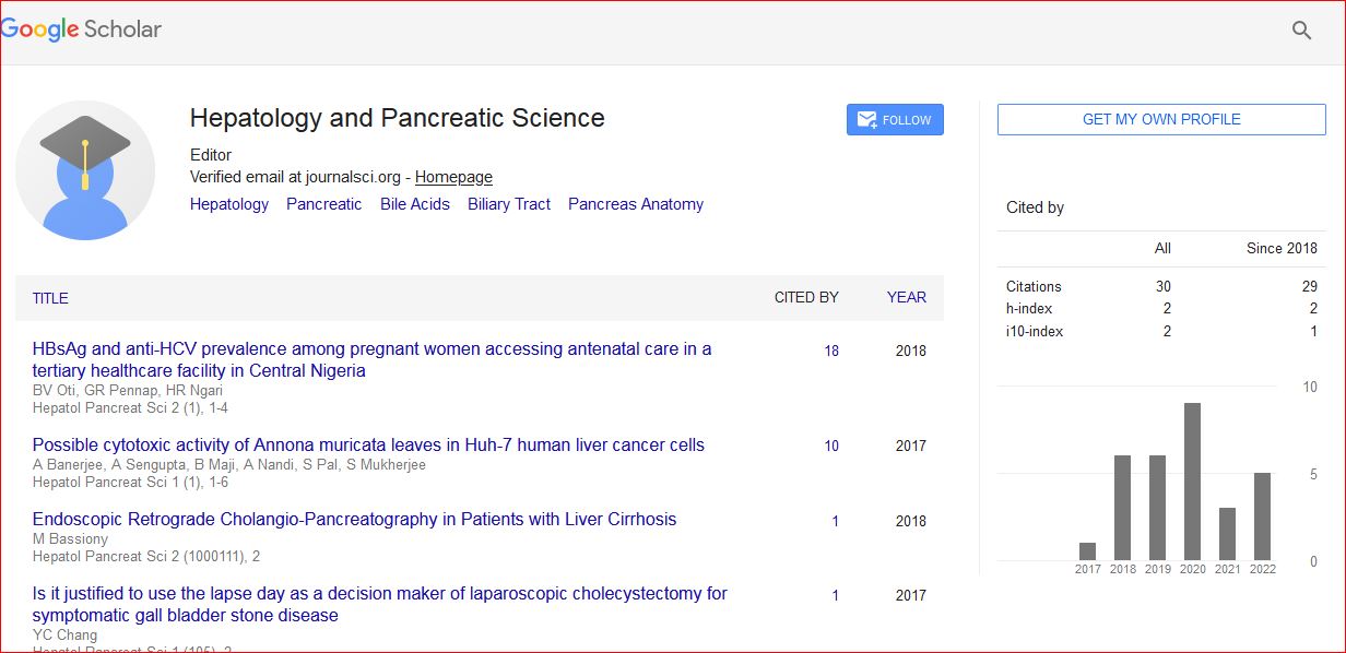
Hepatology and Pancreatic Science received 34 citations as per Google Scholar report