Nayomi Shermila
Treatment of dengue stays supportive in the lack of targeted antiviral therapy or approved vaccines. Responsive fluid management is key to prohibiting progression to shock or other severe manifestations. The dynamic natural history of dengue infection and its influence on hemodynamic homeostasis needs to be carefully considered in the planning of individualized therapy. Though largely self-limiting, the sheer burden of dengue disease on the global population will outcome in a typical manifestations especially in children, older adults, and comorbid patients. Management of these has not yet been systematized. The failure of recent randomized controlled trials to show use for antiviral and immunomodulatory agents in dengue is dejecting. Vaccine candidates have promise, but growing outbreaks need more robust, evidence-based management guidelines to inform clinicians, mainly in novel epidemic situations.
Dengue fever is caused by a flavivirus, which is a vector borne RNA virus with four anti-genically distinct serotypes (DEN 1, DEN 2, DEN 3 and DEN 4). Neurological manifestations are rare compared to other complications of the disease. Encephalopathy, encephalitis, aseptic meningitis, intracranial hemorrhages, thrombosis, mono-neuropathies/polyneuropathies, Guillain-Barre syndrome and myelitis have been reported. Neurological manifestation in dengue hemorrhagic fever usually results from multisystem dysfunction secondary to liver failure, cerebral hypoperfusion, electrolyte imbalance, shock, cerebral edema and hemorrhage related to vascular leak. The occurrence of brain hemorrhage in a case with dengue shock can be serious and leads to death. The occurrence of brainstem hemorrhage can be a very serious fatal situation. We report this case series of dengue hemorrhagic fever with multiple intracranial, sub arachnoid hemorrhages and sub-dural hematoma causing brainstem herniation. Case 1: A 25-year-old previously healthy woman was admitted on third day of fever with thrombocytopenia. Critical phase started on 5th day with evidence of pleural effusion and moderate ascites. 31 hours into critical phase, she developed headache, altered level of consciousness, limb rigidity and respiratory depression without definite seizures. Non-contrast CT brain done at tertiary care level revealed diffuse intra cranial hemorrhages and sub arachnoid hemorrhages in right frontal, parietal, occipital lobes and brainstem, cerebral oedema with an acute subdural hematoma in right temporo-parietal region. Her platelet count was 40,000 at this time with signs of vascular leakage. She was intubated and ventilated with supportive care. Later on, she developed features of cranial diabetes insipidus and it responded to intranasal desmopressin therapy. In spite of above measures signs of brainstem herniation developed and she succumbed to the illness on day 8. Dengue was confirmed serologically. Case 2: A 24 year old previously healthy was admitted on 2nd day of fever with constitutional symptoms and no bleeding manifestations. Clinical, hematological and serological parameters confirmed dengue infection. On 5th day of illness, she entered into leaking phase, but did not have evidence of any bleeding Intra Cranial Hemorrhage (ICH) in right parietal lobe deep white matter area associated with perilesional oedema and midline shift. Bleeding into the right lateral ventricle and Small Subdural Hematoma (SDH) were also noted in right parietal lobe area. Her platelet count at the time of development of hemorrhages was 32,000 and International Normalised Ratio was normal. NCCT brain was repeated 24 hours later and showed progression of hemorrhages. It showed progressive worsening of right occipito-temporal ICH, cerebral oedema, midline shift, right SDH and SAH. Patient remained hemodynamically stable and platelet count was on the rising trend. It was 52,000, 77,000 and 83,000 on 3 consecutive occasions. PCV was stable around 43. There were no other bleeding manifestations neurosurgical interventions were not tried and patient was treated conservatively. With maximum care provided, patient succumbed to illness on the following day. It can be concluded that diffused cerebral hemorrhages with moderate thrombocytopenia and normal coagulation profile are a very rare and fatal complication of dengue fever. Exact pathophysiological mechanism is not well understood. Increased awareness and high degree of clinical suspicion is needed among clinicians for timely diagnosis of this extremely rare complication of dengue fever. We postulate that immunological mechanisms may play a role in pathogenesis. However, more comprehensive research and analysis are needed to understand the pathophysiological mechanisms leading to this complication.
The impact of dengue has the potential for severe morbidity in young and healthy individuals, especially in situations of massive outbreaks, as have been happening, for example, in Lahore, Pakistan in 2011, with 500,000 notified cases in a city of 5 million, and a dengue IgGseropositivity rate of 67.9 % the following year in a city not previously endemic for dengue
PDFShare this article
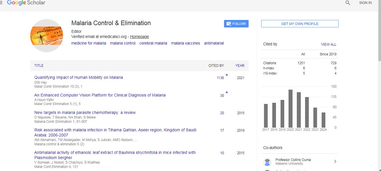
Malaria Control & Elimination received 1187 citations as per Google Scholar report