Madhumita Das, Bishwajit Das, Avishek Das, Ishita Chattrejee and Durjoy Majumder
Light microscopic images are routinely used in clinical diagnosis. Further the confirmation of disease diagnosis is relied with the ultra-structural images. Scanning electron microscopy (SEM) with high resolution and high magnification is granted as one of the reliable process in this aspect. However, pathologists’ criteria for a disease diagnosis for both the processes are on the basis of qualitative and empirical in nature. In a number of hematological diseases as well as in different pathological conditions like liver cirrhosis morphological alteration of red blood cell alteration is reported. Recently some morphological alteration in red blood cells (RBC) by ultra-structural analysis has been reported in leukemic patients, though leukemia is a disease of white blood cells. It is expected that these features could be used as an event of cancer cachexia or can be used as the identifying marker for pre-cancer state. However, a systematic study so far is not done in terms of proper quantitative statement. Here an attempt has been made towards the development of an automated image analysis procedure for extracting quantitative information from the scanning electron microscopic (SEM) images. This computational approach may guide the clinicians to take a decision about the disease/pre-disease state with a quantitative outlook.
PDFShare this article
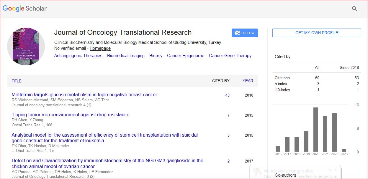
Journal of Oncology Translational Research received 93 citations as per Google Scholar report